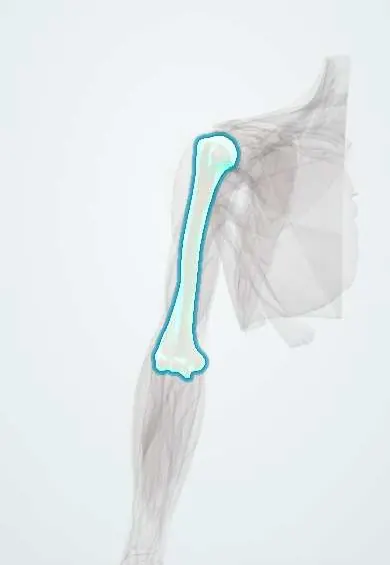
Supracondylar humerus fracture !
A supracondylar humerus fracture is a break just above the elbow, the most common elbow fracture in children; it can injure the artery and nerves nearby.
Updated 2026-10-29
Structures involved
Tap one to see what it is and open it in 3D.
- Humerus
- Brachial artery
- Median nerve
- Anterior interosseous nerve
- Radial nerve
- Ulnar nerve
- Brachialis
- Elbow capsule
- FDP
- FPL
In the clinical exam
Why it happens
What it is
Fracture of the distal humerus just above the condyles, the most common elbow fracture in children. Most are extension type, and the displaced fragment can injure the brachial artery and median or anterior interosseous nerve.
Who gets it
Mainly young children, typically between about 5 and 7 years, from falls from playground equipment, beds, trees or bicycles. It is a frequent paediatric orthopaedic emergency in Colombian hospitals.
Causes
- Fall on an outstretched hand with the elbow hyperextended (extension type, by far the most common)
- Fall directly on a flexed elbow (flexion type, uncommon)
Risk factors
- Age of about 5 to 7 years
- Falls from heights: playground equipment, trampolines, bunk beds, trees
- Ligamentous laxity allowing elbow hyperextension in children
Pathophysiology
In a child the thin supracondylar bone, between the olecranon and coronoid fossae, is weaker than the ligaments. In hyperextension the olecranon levers into its fossa and the humerus breaks; the distal fragment moves backwards and the sharp proximal end pushes forwards toward the brachial artery and median nerve. Swelling can cause forearm compartment syndrome.
Classification
- Extension type (most common) or flexion type
- Gartland (extension type): I non-displaced; II displaced with intact posterior cortex; III completely displaced; IV unstable in flexion and extension
- Posterolateral displacement threatens the brachial artery and median nerve; posteromedial threatens the radial nerve
Symptoms and signs
- Elbow pain, swelling and refusal to use the arm after a fall
- S-shaped deformity of the elbow in displaced fractures
- Arm held in extension or slight flexion
- Bruising or skin puckering in front of the elbow (anterior spike)
- Loss of the OK sign (thumb and index flexion) if the anterior interosseous nerve is injured
Diagnosis
- Clinical
- Careful documented neurovascular examination before any treatment: radial pulse, capillary refill, hand colour, and anterior interosseous, median, radial and ulnar nerve function.
- Tests
- Usually not needed; pulse oximetry or Doppler may support the vascular assessment.
- Imaging
- AP and lateral elbow X-rays: posterior fat pad sign, anterior humeral line not crossing the middle third of the capitellum, and Baumann angle; comparison views sometimes.
Differential diagnosis
- Lateral condyle fracture of the humerus
- Medial epicondyle fracture
- Elbow dislocation
- Radial head subluxation (pulled elbow)
- Non-accidental injury in a young child
Goal of treatment
Restore alignment and stable fixation, preserve circulation and nerve function, and prevent compartment syndrome, Volkmann contracture and cubitus varus.
Treatment
- Acute
- Splinting in a comfortable position without forced flexion, analgesia, and repeated neurovascular checks; urgent reduction if the hand is pale and pulseless.
- Conservative
- Type I (and some stable type II) fractures: above-elbow cast for a few weeks.
- Surgical
- Displaced fractures: closed reduction and percutaneous pinning under anaesthesia; open reduction if irreducible or if the artery needs exploration.
- Follow-up
- Postoperative checks of circulation, nerve function and forearm compartment pressure signs.
Complications
- Volkmann ischaemic contracture after untreated compartment syndrome
- Brachial artery injury
- Nerve palsy (anterior interosseous most often; also radial and ulnar, including iatrogenic ulnar injury from a medial pin)
- Cubitus varus (gunstock deformity) from malunion
- Elbow stiffness and pin-site infection
Prognosis
With timely reduction and fixation most children recover full function, and most nerve injuries recover spontaneously. Missed compartment syndrome can cause permanent, disabling contracture of the forearm and hand.
Prevention
- Supervision of young children on playground equipment and trampolines
- Safety rails on bunk beds
Test yourself
Think of the answer, then tap the question to see it.
Which nerve is most often injured in extension-type supracondylar fractures?
The anterior interosseous nerve, a branch of the median nerve: the child cannot make the OK sign.
Which artery is at risk in a displaced supracondylar humerus fracture?
The brachial artery, pushed by the sharp proximal fragment in front of the elbow.
What is Volkmann ischaemic contracture?
Fibrosis and shortening of necrotic forearm flexor muscles after untreated compartment syndrome, causing a clawed, flexed wrist and fingers.
What does the Gartland classification describe?
Displacement of extension-type fractures: I non-displaced, II posterior cortex intact, III completely displaced.
What deformity results from malunion of a supracondylar fracture?
Cubitus varus, also called gunstock deformity.
Related conditions
- Elbow dislocation
- Radial head subluxation (pulled elbow)
- Humeral shaft fracture
- Volkmann ischaemic contracture
Educational information, not medical advice. For any symptom, see a health professional.
Sources: Supracondylar Humerus Fractures (StatPearls (NCBI Bookshelf), 2023).
Spotted a mistake?We are always improving for the community. Tell us what is wrong and our team will review it.Report a mistake
Representative model, for educational purposes only.