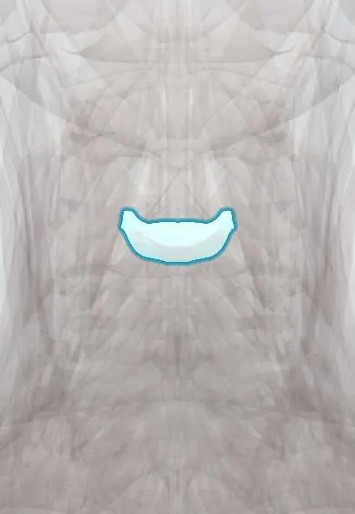
C3–C4 disc !
The C3-C4 disc is the cushion between the C3 and C4 neck vertebrae; it absorbs load and lets the neck bend and turn.
Official name: C3-C4
Opens in the Durgical app, no sign-up needed.
What it is
The cushion between C3 and C4: a tough outer ring of fibres (annulus fibrosus) around a soft centre.
What it does
Absorbs load and lets the neck bend and turn. C5-C6 is the most mobile and most worn neck level.
Key facts
- Where
- Between C3 and C4 bodies; taller in front (cervical lordosis); flanked by uncovertebral joints
- Attaches
- Annulus fibrosus anchored to the endplates and ring apophyses of C3 and C4; anterior and posterior longitudinal ligaments
- Function
- Absorbs and spreads load; posterolateral herniation usually compresses the C4 root
Relations
- In front
- Longus colli and prevertebral fascia; in front, the lower pharynx and the larynx, with the hyoid and the upper border of the thyroid cartilage near this level
- Behind
- Posterior longitudinal ligament, epidural veins, the dural sac and the spinal cord
- Lateral
- Uncovertebral joints, the vertebral artery in the transverse foramina and the C4 root leaving through the C3–C4 foramen
How to find it
From the front, the C3–C4 disc lies near the hyoid and the upper edge of the thyroid cartilage. From behind it is between the C3 vertebra and C4 vertebra spinous processes, which are short and hard to tell apart under the neck muscles.
Examination
- Hoffmann sign: flicking the nail of the middle finger makes the thumb and index finger flex; a positive sign suggests an upper motor neuron problem such as cord compression in the neck
- Lhermitte sign: an electric feeling down the spine or into the limbs when the neck bends forward; it points to a problem in the cord, such as compression
On imaging
Lateral cervical radiographs show disc height and osteophytes; after trauma, a widened soft-tissue shadow in front of the upper cervical spine suggests haematoma. MRI is the test of choice: sagittal T2 shows the disc pressing on the cord and, in myelopathy, high signal inside the cord. In older people with spondylosis, a hyperextension injury may leave a normal CT, so MRI looks for cord damage.
Common problems
- Herniation pressing on the C6 nerve root: neck pain and pain, numbness or weakness down the arm into the thumb side of the hand.
- Degeneration with bone spurs, which can narrow the canal and press on the spinal cord (myelopathy: clumsy hands, unsteady walking).
How it is diagnosed
Exam of arm strength, reflexes and feeling, plus signs of cord pressure. MRI shows the disc, nerve roots and cord.
Treatment
- Most arm pain from a disc improves with time, physical therapy, medicine and sometimes an injection.
- Lasting arm pain, weakness or cord pressure: anterior cervical discectomy and fusion (ACDF), or disc replacement in selected patients.
Clinical notes
- The anterior approach at C3–C4 passes close to the superior thyroid artery and the superior laryngeal nerve; injury to its external branch can lower the voice's range
- C4 shoulder-top pain is easily confused with a trapezius strain or with shoulder disease
- After anterior cervical surgery, a collection of blood in the space in front of the spine can squeeze the airway within hours; the risk is higher with upper and multilevel operations, so breathing and neck swelling are watched closely
Development
Like every disc, it forms from mesenchyme between the vertebral bodies; remnants of the notochord persist in its centre and contribute to the nucleus pulposus, while the rest of the notochord disappears inside the bodies.
Test yourself
Think of the answer, then tap the question to see it.
What is the clinical picture of the C4 root compressed by a C3–C4 disc herniation?
Pain and numbness over the base of the neck and the top of the shoulder (cape area), with no reliable reflex or single muscle to test.
How is the Hoffmann sign elicited, and what does it suggest in cord compression at C3–C4?
Flicking the nail of the middle finger makes the thumb and index finger flex; it suggests an upper motor neuron problem such as cervical cord compression.
Which nerve is at risk in an anterior approach to the C3–C4 disc, and what does its injury cause?
The superior laryngeal nerve, near the superior thyroid artery; injury to its external branch can lower the voice's range.
Educational information, not medical advice. For any symptom, see a health professional.
Its names
- Official Latin (TA2)
- discus intervertebralis C3-C4 TA2 1684
- Official Spanish (DeCS)
- Disco C3-C4 DeCS D007403
- English (TA2)
- C3-C4
- Also called
- fibrous ring · fibrocartilago intervertebralis
Related
Sources: Terminologia Anatomica (TA2); DeCS; Anatomy, Back, Intervertebral Discs; 7.5 Embryonic Development of the Axial Skeleton (OpenStax (Rice University), 2022) · adapted, CC BY 4.0.
Spotted a mistake?We are always improving for the community. Tell us what is wrong and our team will review it.Report a mistake
Representative model, for educational purposes only.