Joints and ligaments · Lower limb
Toe joint capsules !
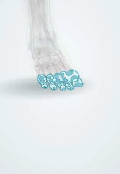
The toe joint capsules are sleeves around the joints of the toes that hold the joint fluid.
Official name: Capsules of metatarsophalangeal and interphalangeal joints of foot
Opens in the Durgical app, no sign-up needed.
What it is
Thin sleeves of tissue around the joints at the base of the toes and inside the toes.
What it does
Enclose each joint and hold the fluid that lubricates it.
Key facts
- Where
- Metatarsophalangeal and interphalangeal joints of the toes
- Attaches
- Articular margins of metatarsal heads and phalanges; reinforced by collateral ligaments and plantar plates
- Function
- Enclose synovial joints; blend dorsally with extensor expansions
Relations
- Above
- Extensor tendons and their hoods (extensor expansions), blended with the dorsal capsule
- Below
- Plantar plates with the flexor tendons in their fibrous sheaths on them; at the big toe, the two sesamoids
- Lateral
- On each side, the collateral ligaments; between neighbouring joints, the deep transverse metatarsal ligament links the plantar plates, with the lumbricals and digital nerves passing below it
How to find it
The metatarsophalangeal joint lines are felt on the top of the foot just beyond the metatarsal heads when the toes are passively bent down; synovitis makes them boggy and tender. The interphalangeal joints are felt as the knuckles of each toe.
Examination
- Vertical stress (drawer) test of a lesser toe: lifting the proximal phalanx upward on the metatarsal head; excess movement or pain suggests a plantar plate tear
- Metatarsophalangeal squeeze test: pain on squeezing the forefoot from side to side suggests MTP synovitis, as in early rheumatoid arthritis
On imaging
Weight-bearing radiographs show joint space, toe alignment, sesamoid position and erosions (punched-out with overhanging edges in gout, marginal in rheumatoid arthritis). Ultrasound shows effusion, synovitis and plantar plate tears; MRI shows occult injuries.
Common problems
- Swelling and pain with arthritis, gout (often the big toe) or injury.
Clinical notes
- Rheumatoid arthritis often begins in the MTP joints; stretched capsules and failed plantar plates lead to clawed, dorsally dislocated toes with prominent metatarsal heads
- Hallux valgus surgery often combines a lateral capsular release with tightening of the medial capsule
- Hallux rigidus: degenerative disease of the first MTP joint with a thickened capsule and dorsal osteophytes that block toe extension
Anatomical variants
- A bipartite sesamoid, most often the medial (tibial) one, is a common variant that can be mistaken for a fracture
Development
Each capsule forms from the mesenchyme around the gap between neighbouring cartilage models, where the joint cavity opens; thicker parts of the capsule become the collateral ligaments and plantar plates.
Related conditions
Test yourself
Think of the answer, then tap the question to see it.
What forms the plantar side of each metatarsophalangeal joint capsule?
The plantar plate, a thick fibrocartilage; at the big toe it holds the two sesamoids.
Which arthritis often begins in the MTP joint capsules and leads to clawed, dorsally dislocated toes?
Rheumatoid arthritis; the MTP squeeze test is painful in early disease.
What is hallux rigidus?
Degenerative disease of the first MTP joint, with a thickened capsule and dorsal osteophytes that block big-toe extension.
Educational information, not medical advice. For any symptom, see a health professional.
Its names
- Official Latin (TA2)
- articulationes metatarsophalangeae; articulationes interphalangeae pedis TA2 1964
- Official Spanish (DeCS)
- Cápsulas de las articulaciones metatarsofalángicas e interfalángicas del pie DeCS D017746
- English (TA2)
- Capsules of metatarsophalangeal and interphalangeal joints of foot
- Also called
- articuli metatarsophalangici
Related
- MTP ligaments and plantar plate
- Toe ligaments
- First metatarsal
- Second metatarsal
- Third metatarsal
- Fourth metatarsal
- Fifth metatarsal
- Big toe
Sources: Terminologia Anatomica (TA2); DeCS; Anatomy, Bony Pelvis and Lower Limb, Foot Joints; 9.7 Development of Joints (OpenStax (Rice University), 2022) · adapted, CC BY 4.0; Anatomy of the Human Body (20th ed.): Development of the Joints (Lea & Febiger (Gray's Anatomy, 1918), 1918) · adapted, Public domain.
Spotted a mistake?We are always improving for the community. Tell us what is wrong and our team will review it.Report a mistake
Representative model, for educational purposes only.