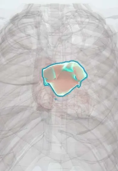
Cardiac tumours (atrial myxoma) !
A benign, jelly-like tumour inside the heart, usually in the left atrium. It can block blood flow or shed clots and is removed by surgery.
Updated 2026-11-01
Structures involved
Tap one to see what it is and open it in 3D.
- Left atrium
- Interatrial septum
- Mitral anterior leaflet
- Mitral posterior leaflet
- Right atrium
- Left ventricle
- Pulmonary veins
Why it happens
- Benign vs malignant tumours: the comparison table
- Systemic (arterial) thromboembolism
- Murmurs and the hemodynamics of valve disease
What it is
Primary cardiac tumours are rare; metastases to the heart are far more common. Myxoma is the most common primary cardiac tumour in adults: a soft, gelatinous, usually pedunculated mass, most often in the left atrium attached by a stalk to the interatrial septum near the fossa ovalis. Though benign, it can obstruct the mitral valve, embolise and cause systemic inflammatory symptoms.
Who gets it
Primary cardiac tumours are found in well under 0.1% of autopsies, and about half of benign primary tumours in adults are myxomas. Myxomas are most common in women between 30 and 60 years. About 7–10% are familial, most often as part of Carney complex. In children the most common primary cardiac tumour is rhabdomyoma, linked to tuberous sclerosis.
Causes
- Sporadic myxoma: arises from multipotent mesenchymal cells in the endocardium, usually near the fossa ovalis
- Familial myxoma in Carney complex: autosomal dominant, usually variants in PRKAR1A
- Other primary tumours: papillary fibroelastoma (valves), rhabdomyoma and fibroma (children), lipoma, angiosarcoma (most common primary malignant tumour)
- Secondary tumours (most common overall): metastases from lung, breast, melanoma, lymphoma and leukaemia
Risk factors
- Female sex and middle age (sporadic myxoma)
- Family history of myxoma or Carney complex
- Tuberous sclerosis (rhabdomyoma in infants)
- Advanced cancer (cardiac metastases)
Pathophysiology
Myxomas cause disease by three mechanisms. Obstruction: a mobile left atrial tumour prolapses into the mitral orifice in diastole, mimicking mitral stenosis and raising left atrial and pulmonary pressures; symptoms may change with posture. Embolism: friable fragments or overlying thrombus travel to the brain, limbs, kidneys or coronary arteries (right-sided tumours embolise to the lungs). Constitutional effects: the tumour secretes interleukin-6, causing fever, weight loss, arthralgia, anaemia and raised inflammatory markers.
Classification
- Primary benign: myxoma, papillary fibroelastoma, rhabdomyoma, fibroma, lipoma
- Primary malignant: sarcomas (angiosarcoma most common), primary cardiac lymphoma
- Secondary: metastases by direct, lymphatic or haematogenous spread
- Myxoma by location: left atrium (about 75%), right atrium, ventricles (more often in Carney complex)
Symptoms and signs
- Dyspnoea, orthopnoea and pulmonary oedema (mitral obstruction)
- Dizziness or syncope, sometimes positional
- Sudden neurological deficit or limb ischaemia (embolism)
- Fever, weight loss, fatigue and arthralgia
- May be asymptomatic and found incidentally on echocardiography
Diagnosis
- Clinical
- Signs resembling mitral stenosis that vary with position; an early diastolic «tumour plop» after S2; constitutional symptoms. In Carney complex look for spotty skin pigmentation (lentigines), cutaneous myxomas and endocrine overactivity (Cushing syndrome, acromegaly).
- Tests
- Raised erythrocyte sedimentation rate and C-reactive protein, anaemia, hypergammaglobulinaemia. ECG is nonspecific. Histology after excision confirms the diagnosis; percutaneous biopsy is avoided because of embolic risk. Genetic testing and family screening when Carney complex is suspected.
- Imaging
- Transthoracic echocardiography is the first test; transoesophageal echocardiography defines the attachment site and size. Cardiac MRI characterises tissue and separates tumour from thrombus; CT helps plan surgery and look for metastatic disease.
Differential diagnosis
- Left atrial thrombus (usually in the appendage, with atrial fibrillation or mitral stenosis)
- Rheumatic mitral stenosis
- Infective endocarditis with large vegetations (fever, emboli)
- Other cardiac tumours, including metastases
- Vasculitis or connective tissue disease (constitutional symptoms)
Goal of treatment
Remove the tumour promptly to prevent embolism, obstruction and sudden death, then follow for recurrence.
Treatment
- Surgical
- Prompt surgical excision on cardiopulmonary bypass, removing the stalk with a margin of the interatrial septum and repairing the defect with a patch; the valve is repaired if damaged.
- Acute
- Emergency management of complications while surgery is arranged: stroke or limb ischaemia pathways, treatment of pulmonary oedema; surgery is not delayed once the diagnosis is made.
- Follow-up
- Echocardiographic follow-up for recurrence (more frequent in familial and multiple tumours); screening of first-degree relatives in Carney complex. Nursing after surgery: rhythm monitoring for atrial arrhythmias, neurological checks and wound care.
Complications
- Ischaemic stroke and other systemic emboli
- Sudden death from complete obstruction of the mitral orifice
- Heart failure and pulmonary hypertension
- Arrhythmias
- Recurrence after excision, mainly in Carney complex
Prognosis
After complete excision of a sporadic myxoma the prognosis is excellent and recurrence is uncommon. Familial myxomas recur more often and need lifelong surveillance. Malignant primary tumours and metastases have a poor prognosis.
Prevention
- No primary prevention; family screening and surveillance in Carney complex
- Early echocardiography in young patients with unexplained stroke or emboli
Test yourself
Think of the answer, then tap the question to see it.
Where is the classic atrial myxoma located?
In the left atrium, attached by a stalk to the interatrial septum near the fossa ovalis.
What auscultatory sound is described with a mobile myxoma?
An early diastolic «tumour plop».
Which cytokine produced by myxomas explains fever and raised inflammatory markers?
Interleukin-6.
What is the most common cardiac tumour overall?
Metastasis to the heart.
What is the most common primary cardiac tumour in children, and with which syndrome?
Rhabdomyoma, associated with tuberous sclerosis.
Related conditions
- Mitral stenosis
- Ischaemic stroke
- Transient ischaemic attack
- Syncope (vasovagal, orthostatic hypotension, cardiac)
- Heart failure
- Atrial fibrillation
- Infective endocarditis
- Pulmonary embolism
- Acute limb ischaemia
- Cushing syndrome
- Pulmonary hypertension and cor pulmonale
Educational information, not medical advice. For any symptom, see a health professional.
Sources: Atrial myxoma (MedlinePlus, US National Library of Medicine, 2026); Carney complex (MedlinePlus Genetics, US National Library of Medicine, 2026).
Spotted a mistake?We are always improving for the community. Tell us what is wrong and our team will review it.Report a mistake
Representative model, for educational purposes only.