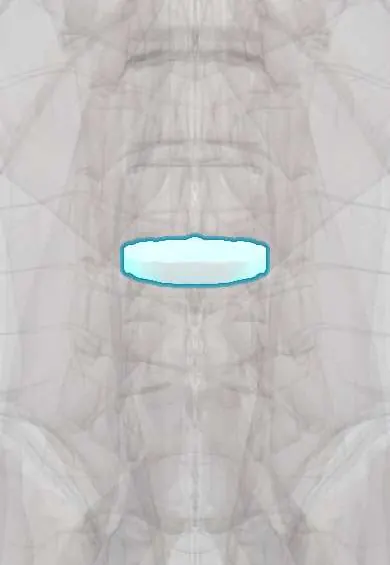
L2–L3 disc !
The L2-L3 disc is the cushion between the L2 and L3 vertebrae in the lower back; it absorbs shock and lets the spine bend and twist.
Official name: L2-L3
Opens in the Durgical app, no sign-up needed.
What it is
The cushion between L2 and L3: a tough outer ring of fibres (annulus fibrosus) around a soft centre.
What it does
Absorbs shock, spreads load across the vertebrae and lets the spine bend and twist, while holding the bones together.
Key facts
- Where
- Between L2 and L3 bodies; thick lumbar disc, taller in front (lumbar lordosis)
- Attaches
- Annulus fibrosus anchored to the endplates and ring apophyses of L2 and L3; anterior and posterior longitudinal ligaments
- Function
- Absorbs and spreads load; posterolateral herniation usually compresses the L3 root
Relations
- In front
- Abdominal aorta and inferior vena cava, with the right crus of the diaphragm reaching down to this level
- Behind
- Posterior longitudinal ligament and the dural sac holding the cauda equina, below the end of the cord
- Lateral
- Psoas major with the lumbar plexus inside it; the L2 root exits through the foramen at this level while the L3 root passes down behind the disc
How to find it
Usually about two levels above the Tuffier line. Levels are counted on X-ray, keeping in mind that a transitional lumbosacral vertebra can shift the count by one.
Examination
- Rising from a chair or a step-up on one leg: shows mild quadriceps weakness that manual testing can miss
- Femoral nerve stretch test with the person prone: front-of-thigh pain when the knee is bent supports L2–L3 or L3–L4 root irritation
On imaging
MRI is the test of choice. On axial T2 a paracentral herniation at L2–L3 presses on the traversing L3 root, while a foraminal or far-lateral one presses on the exiting L2 root; sagittal images through the foramina help. Radiographs show narrowing and spurs. A far-lateral herniation is easy to miss if the axial slices do not include the foramina.
Common problems
- Herniation (slipped disc): the gel pushes through a tear in the ring and presses on a nerve root, causing sciatica down the leg.
- Degeneration with age: the disc dries out, loses height and can bulge, sometimes with back pain.
- Tears in the ring (annular fissures) that can cause back pain.
How it is diagnosed
History and exam (the straight-leg raise test, strength, reflexes and feeling in the leg). MRI shows the disc and which nerve is pressed; it is usually done only if pain lasts more than about 6 weeks or there are warning signs.
Treatment
- Most herniations improve within weeks to months: staying active, physical therapy, pain medicine, sometimes an epidural steroid injection.
- Severe or lasting leg pain, or weakness: microdiscectomy to remove the herniated piece.
- Urgent surgery if there are signs of cauda equina syndrome (numbness in the saddle area, bladder or bowel changes).
- Selected cases of disc collapse with instability: fusion with a cage and bone graft.
Clinical notes
- Lateral (transpsoas) approach for interbody fusion at L2–L3: the lumbar plexus lies in the back of the psoas major and the genitofemoral nerve on its front, so nerve monitoring is used
- Diabetic amyotrophy also weakens the thigh with pain, but usually without the back pain and dermatomal pattern of an L3 root lesion
Development
The lumbar curve is a secondary curve that appears as the child begins to stand and walk. The lumbar discs are deeper in front than behind and share in shaping it, together with the wedge of the lowest bodies.
Test yourself
Think of the answer, then tap the question to see it.
What is the L3 root picture from an L2–L3 disc herniation?
Weak knee extension (quadriceps) and hip adduction, a reduced knee jerk (shared with L4), and numbness of the lower front of the thigh to the inner knee.
How is an L3 root lesion from the L2–L3 disc told apart from a femoral nerve lesion?
By hip adduction: it is weak in an L3 root lesion but spared in a femoral nerve lesion, because the obturator nerve supplies the adductors.
At the L2–L3 disc, which root exits through the foramen and which passes behind the disc?
The L2 root exits through the foramen at this level; the L3 root passes down behind the disc.
Which nerves are at risk in a lateral transpsoas fusion at the L2–L3 disc?
The lumbar plexus in the back of the psoas major and the genitofemoral nerve on its front, so nerve monitoring is used.
Educational information, not medical advice. For any symptom, see a health professional.
Its names
- Official Latin (TA2)
- discus intervertebralis L2-L3 TA2 1684
- Official Spanish (DeCS)
- Disco L2-L3 DeCS D007403
- English (TA2)
- L2-L3
- Also called
- fibrous ring · fibrocartilago intervertebralis
Related
Sources: Terminologia Anatomica (TA2); DeCS; Anatomy, Back, Intervertebral Discs; 7.3 The Vertebral Column (OpenStax (Rice University), 2022) · adapted, CC BY 4.0; Anatomy of the Human Body (20th ed.): Articulations of the Vertebral Column (Lea & Febiger (Gray's Anatomy, 1918), 1918) · adapted, Public domain.
Spotted a mistake?We are always improving for the community. Tell us what is wrong and our team will review it.Report a mistake
Representative model, for educational purposes only.