
Appendiceal neoplasms: neuroendocrine tumour, mucinous neoplasms and mucocele, adenocarcinoma
About 1 in 100 removed appendices holds a tumour, usually a surprise on the pathology report. Size, site and type decide whether more surgery is needed.
What it is
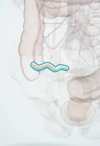
Tumours of the appendix are rare and are most often found by chance in an appendix removed for appendicitis. The main groups are neuroendocrine tumours (formerly called carcinoids), usually small and at the tip; mucinous neoplasms (low-grade and high-grade appendiceal mucinous neoplasms, LAMN and HAMN), which can swell the appendix into a mucus-filled 'mucocele' and, if they rupture, cause pseudomyxoma peritonei; and adenocarcinoma, including colonic-type, mucinous and goblet cell adenocarcinoma, which behaves like colon cancer.
Who gets it
Appendiceal tumours are found in roughly 0.5–1% of appendicectomy specimens. Neuroendocrine tumours are usually found in younger adults, often in their thirties or forties, and are slightly commoner in women; mucinous neoplasms and adenocarcinoma peak in the fifties and sixties. The risk of an underlying tumour rises in patients over about 40 with complicated appendicitis or a periappendiceal abscess, which matters in Colombia where many patients present late and are first managed without surgery.
Causes
- Neuroendocrine tumour: arises from neuroendocrine cells of the appendiceal lining; cause unknown
- Mucinous neoplasms: epithelial tumours that make mucin and push it into a blocked lumen
- Adenocarcinoma: like colorectal cancer, from serrated or adenomatous precursors in some
- Non-neoplastic mucocele: simple retention cyst from obstruction of the lumen
Risk factors
- Older age (mucinous neoplasms and adenocarcinoma)
- Complicated appendicitis with abscess or phlegmon in an adult over about 40
- Appendix diameter above about 15 mm on imaging
- Personal history of colorectal neoplasia (synchronous colon tumours occur with mucinous lesions)
- Hereditary syndromes such as multiple endocrine neoplasia type 1 (rare, for neuroendocrine tumours)
Pathophysiology
Neuroendocrine tumour: grows as a small yellow nodule, usually at the tip, so it rarely obstructs the lumen early → it is found by chance after appendicectomy → risk of nodal spread rises with size, base location, invasion of the mesoappendix and higher grade → spread to the liver is rare, and only then can serotonin reach the systemic circulation and cause carcinoid syndrome. Mucinous neoplasm: tumour lines the lumen and keeps making mucin → the appendix distends into a mucocele, sometimes with calcified walls → pressure thins the wall → it ruptures or leaks → mucin and tumour cells seed the peritoneum (pseudomyxoma peritonei). Adenocarcinoma: invades the wall and spreads to lymph nodes, liver and peritoneum like colon cancer.
Classification
- Neuroendocrine tumour (NET): grade 1–3 by Ki-67 and mitoses; size bands under 1 cm, 1–2 cm and over 2 cm
- Mucinous: LAMN (low-grade, pushing border) and HAMN (high-grade cytology, no infiltrative invasion)
- Mucocele: a descriptive term for a mucus-distended appendix; the cause may be a retention cyst, serrated lesion, LAMN or mucinous adenocarcinoma
- Adenocarcinoma: colonic-type, mucinous, signet-ring and goblet cell adenocarcinoma (formerly goblet cell carcinoid)
- Rare: lymphoma and metastases to the appendix
Symptoms and signs
- Most are found incidentally, in an appendix removed for appendicitis or on imaging for another reason
- Right lower quadrant pain identical to acute appendicitis (from obstruction of the lumen)
- Palpable right iliac fossa mass (large mucocele)
- Abdominal swelling from pseudomyxoma peritonei after rupture
- Flushing, diarrhoea and wheeze (carcinoid syndrome) only with liver metastases, and rare
Diagnosis
- Clinical
- Most diagnoses come from the pathology report after appendicectomy; read it for type, size, site (tip or base), margin, grade, mesoappendiceal invasion, lymphovascular invasion and perforation. In an adult with an appendiceal mass or a dilated appendix on imaging, think mucinous neoplasm before planning surgery.
- Tests
- Histology with immunohistochemistry (chromogranin and synaptophysin for neuroendocrine tumour; Ki-67 for grade). CEA, CA 19-9 and CA-125 for mucinous tumours and adenocarcinoma. Chromogranin A and urinary 5-HIAA only when metastatic neuroendocrine disease or carcinoid syndrome is suspected. Colonoscopy for mucinous lesions and adenocarcinoma, looking for synchronous colorectal neoplasms.
- Imaging
- CT of the abdomen and pelvis: a dilated (over about 15 mm), fluid-filled tubular appendix with a thin, sometimes curvilinear calcified wall suggests a mucocele; a soft-tissue mass suggests adenocarcinoma; check for free mucin and peritoneal spread. Ultrasound may show an onion-skin layered appearance of a mucocele. For neuroendocrine tumour over 2 cm or high risk: CT or MRI for staging and somatostatin-receptor PET-CT for distant disease.
Differential diagnosis
- Acute appendicitis without tumour
- Appendiceal abscess or phlegmon
- Caecal carcinoma
- Crohn disease of the terminal ileum and appendix
- Ovarian cyst or mucinous ovarian tumour
- Small bowel neuroendocrine tumour of the terminal ileum
Tumour in the appendicectomy report: what next?
| Finding | Usual site and look | How it spreads | Usual next step |
|---|---|---|---|
| Neuroendocrine tumour under 2 cm at the tip, low grade, no adverse features | Small yellow nodule at the tip | Almost never | Appendicectomy is enough; no routine follow-up |
| Neuroendocrine tumour over 2 cm, at the base, or with adverse features | Larger nodule, base or mesoappendix | Lymph nodes along the ileocolic vessels | Right hemicolectomy and staging |
| Low-grade mucinous neoplasm (LAMN), intact | Distended mucus-filled appendix (mucocele) | Rupture: mucin into the peritoneum | Appendicectomy with clear margin, colonoscopy, imaging follow-up |
| Adenocarcinoma (colonic-type, mucinous, goblet cell) | Mass or thickened wall, may perforate | Nodes, liver and peritoneum | Right hemicolectomy, staging CT, chemotherapy as for colon cancer |
| Acute appendicitis (no tumour) | Inflamed, swollen appendix, often a faecolith | Perforation, abscess | Nothing further |
Goal of treatment
Remove the tumour completely with the smallest operation that cures it, add a right hemicolectomy only when lymph node spread is a real risk, keep any mucinous lesion intact, and refer peritoneal spread to a specialised peritoneal surface centre.
Treatment
- Surgical
- Neuroendocrine tumour under 1 cm with clear margin, and most 1–2 cm tumours without adverse features: appendicectomy alone is curative.
- Surgical
- Neuroendocrine tumour over 2 cm, at the base with involved margin, with deep mesoappendiceal or lymphovascular invasion, or grade 2–3: completion right hemicolectomy to remove the ileocolic lymph nodes.
- Surgical
- Suspected mucocele: careful laparoscopic or open appendicectomy without grasping or rupturing it, removed in a bag, with a clear margin at the base (sometimes a cuff of caecum); no percutaneous biopsy, and the abdomen inspected and any free mucin sampled.
- Surgical
- Adenocarcinoma, including goblet cell adenocarcinoma: right hemicolectomy with lymphadenectomy and staging, as for colon cancer; in women with mucinous tumours the ovaries are inspected.
- Medical
- Adjuvant or palliative systemic chemotherapy for node-positive or metastatic adenocarcinoma; somatostatin analogues for metastatic neuroendocrine tumour and carcinoid syndrome.
- Procedure
- Perforated mucinous neoplasm with pseudomyxoma peritonei: referral for cytoreductive surgery with HIPEC in an experienced centre.
- Follow-up
- LAMN without rupture: periodic CT or MRI and tumour markers for several years; after rupture, closer surveillance even with no visible spread. Colonoscopy for mucinous lesions and adenocarcinoma.
Complications
- Pseudomyxoma peritonei after rupture of a mucinous neoplasm
- Lymph node and liver metastases (adenocarcinoma, larger neuroendocrine tumours)
- Carcinoid syndrome with liver metastases, and carcinoid heart disease (rare)
- Intussusception of the appendix or caecum by a mucocele
- Missed tumour when appendicitis is treated without surgery and not followed up
Common traps
- Every appendiceal neuroendocrine tumour needs a right hemicolectomy: most are small and at the tip, and appendicectomy alone cures them; hemicolectomy is for size over 2 cm or adverse features
- An appendiceal neuroendocrine tumour usually causes carcinoid syndrome: the syndrome needs liver metastases so serotonin bypasses liver breakdown; almost all appendiceal tumours are silent
- A mucocele is a diagnosis: it is only a description of a mucus-filled appendix; the cause ranges from a harmless retention cyst to mucinous adenocarcinoma, and only histology tells
- A dilated appendix can be biopsied with a needle first: puncturing a mucinous neoplasm can seed the peritoneum; it is removed intact instead
- Goblet cell carcinoid is a neuroendocrine tumour: now called goblet cell adenocarcinoma, it behaves like an adenocarcinoma and is treated with right hemicolectomy
Prognosis
Small neuroendocrine tumours at the tip are cured by appendicectomy, with near-normal life expectancy. Intact low-grade mucinous neoplasms removed with clear margins rarely recur; once ruptured they may develop pseudomyxoma over years. Adenocarcinoma behaves like colon cancer of the same stage, and goblet cell and signet-ring types do worse.
Prevention
- Send every appendix for histology
- In adults over about 40 managed without surgery for complicated appendicitis, arrange interval CT and colonoscopy
- Handle a dilated appendix gently and remove it intact
- Follow the pathology report to completion surgery or surveillance when indicated
Test yourself
Think of the answer, then tap the question to see it.
A 28-year-old's appendicectomy report shows a 7 mm grade 1 neuroendocrine tumour at the tip, clear margins, no invasion. What further treatment is needed?
None: appendicectomy is curative for a small, low-grade tumour at the tip without adverse features.
Why does a 2.5 cm neuroendocrine tumour of the appendix base lead to a right hemicolectomy?
Larger and basal tumours have a real risk of spread to the ileocolic lymph nodes and of a positive margin; the right colon is removed with its nodes.
CT in a 60-year-old shows a 3 cm cystic dilated appendix with a calcified wall. What is it, and which surgical rule matters most?
A mucocele, possibly a mucinous neoplasm. Remove it intact, without rupture or needle biopsy, because spilled mucin can seed the peritoneum (pseudomyxoma).
Why is interval colonoscopy and CT advised after non-operative treatment of an appendiceal abscess in a 55-year-old?
Tumours, including mucinous neoplasms and caecal or appendiceal cancer, are more common behind complicated appendicitis in older adults and would otherwise be missed.
Why it happens
Connections
Educational information, not medical advice. For any symptom, see a health professional.
Sources: Appendiceal Tumors (StatPearls (NCBI Bookshelf), 2025); Pseudomyxoma Peritonei (StatPearls (NCBI Bookshelf), 2026).
Spotted a mistake?We are always improving for the community. Tell us what is wrong and our team will review it.Report a mistake
Representative model, for educational purposes only.