
Small bowel tumours: adenocarcinoma, neuroendocrine tumour, lymphoma, GIST and metastases
Tumours of the small bowel are rare and hide beyond the reach of standard scopes, so they surface late as blockage, bleeding or a bowel that telescopes.
Quick facts
- Key signs
- Vague · Nausea · Weight loss and loss of appetite
- Diagnosis
- Histology from biopsy or the resected specimen
Red flags ↓
No doses: the clinician sets them by the current guideline.
What it is
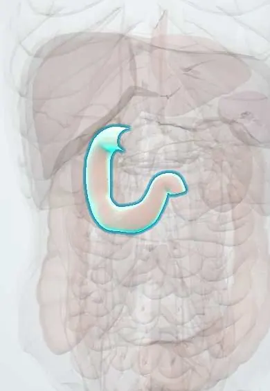
Benign or malignant growths of the duodenum, jejunum or ileum. The five malignant types to know are neuroendocrine tumour (now the commonest, mostly in the ileum), adenocarcinoma (mostly duodenum, often around the ampulla), lymphoma (ileum and jejunum), gastrointestinal stromal tumour (GIST, mostly jejunum and ileum) and metastases (melanoma, lung, breast, or peritoneal spread from ovary and colon). Benign ones include adenomas, lipomas and hamartomatous polyps.
Who gets it
The small bowel makes up most of the length and surface of the gut but fewer than about 5% of gastrointestinal cancers. Incidence rises with age, peaking in the sixties, and has been increasing, mainly because more neuroendocrine tumours are found. Diagnosis is often delayed by months because symptoms are vague. Colombia lacks specific data; regional referral centres see them mainly as obscure bleeding or adult bowel obstruction.
Causes
- Adenocarcinoma: adenoma-to-carcinoma sequence, as in the colon; favoured by inherited polyposis and chronic inflammation
- Neuroendocrine tumour: enterochromaffin cells of the crypts, mostly in the distal ileum; sometimes multiple
- Lymphoma: lymphoid tissue of the bowel wall (B-cell types such as diffuse large B-cell and MALT lymphoma; enteropathy-associated T-cell lymphoma with coeliac disease)
- GIST: interstitial cells of Cajal (the gut pacemaker cells), driven by mutations in KIT or PDGFRA
- Metastases: blood-borne spread (melanoma most typically, also lung and breast) or seeding across the peritoneum
Risk factors
- Familial adenomatous polyposis and Lynch syndrome (adenocarcinoma, especially duodenal)
- Peutz–Jeghers syndrome (hamartomas and adenocarcinoma)
- Crohn disease of the small bowel (adenocarcinoma in inflamed segments)
- Coeliac disease, particularly untreated or refractory (lymphoma and adenocarcinoma)
- Immunosuppression: HIV, transplant drugs (lymphoma)
- Multiple endocrine neoplasia type 1 (duodenal neuroendocrine tumours) and neurofibromatosis type 1 (GIST)
- Previous melanoma
Pathophysiology
A tumour grows in the wall of a long bowel filled with liquid → the liquid flows past it for a long time, so there are few early symptoms → three ways it declares itself: (1) narrowing or acting as a lead point → intermittent colicky pain, intussusception, then obstruction; a neuroendocrine tumour also provokes dense fibrosis in the mesentery that kinks loops and can strangle their blood supply; (2) surface ulceration → slow bleeding → iron deficiency anaemia, or brisk bleeding from a vascular GIST; (3) spread to nodes and liver → in neuroendocrine tumour, amines such as serotonin then bypass breakdown in the liver and reach the circulation → carcinoid syndrome. Periampullary tumours block the bile duct early and cause jaundice; lymphoma can perforate.
Classification
- By cell of origin: epithelial (adenoma, adenocarcinoma), neuroendocrine (well-differentiated tumour, poorly differentiated carcinoma), lymphoid (B-cell and T-cell lymphomas), mesenchymal (GIST, lipoma, leiomyoma, leiomyosarcoma) and metastatic
- By site: duodenum (adenocarcinoma, neuroendocrine tumours), jejunum (GIST, adenocarcinoma, lymphoma), ileum (neuroendocrine tumour, lymphoma)
- Neuroendocrine tumours graded by proliferation (Ki-67 index and mitoses) as G1, G2 or G3
- GIST risk by size, mitotic rate and site; staging of adenocarcinoma by TNM
Symptoms and signs
- Vague, recurrent cramping abdominal pain, often after meals
- Nausea, vomiting and distension from partial or complete obstruction
- Tiredness and pallor from iron deficiency anaemia, or overt black or maroon stools
- Weight loss and loss of appetite
- Jaundice with periampullary tumours
- Flushing, watery diarrhoea and wheeze (carcinoid syndrome) once a neuroendocrine tumour has spread to the liver
Diagnosis
- Clinical
- Suspect it in unexplained iron deficiency or bleeding after negative upper endoscopy and colonoscopy, in adult intussusception and in obstruction without a hernia or previous surgery. Ask about polyposis, coeliac or Crohn disease, immunosuppression and previous melanoma.
- Tests
- Full blood count and iron studies, liver tests and bilirubin. Neuroendocrine tumour: 24-hour urine or plasma 5-hydroxyindoleacetic acid (5-HIAA) and chromogranin A. Carcinoembryonic antigen for adenocarcinoma follow-up. Histology from biopsy or the resected specimen confirms the type (with KIT or DOG1 staining for GIST and markers for lymphoma).
- Imaging
- CT or MR enterography (the bowel distended with oral contrast) shows wall thickening, masses and mesenteric spread; a neuroendocrine tumour typically gives a mesenteric mass with radiating, spiculated strands. Capsule endoscopy views the whole mucosa in patients without strictures; device-assisted (balloon) enteroscopy reaches the lesion to biopsy, treat or tattoo it. Upper endoscopy with a side-viewing scope for duodenal and ampullary lesions. Somatostatin receptor PET-CT stages neuroendocrine tumours; staging CT of chest, abdomen and pelvis for all malignant types.
Differential diagnosis
- Crohn disease stricture
- Adhesive small bowel obstruction
- Small bowel angiodysplasia or Meckel diverticulum (for bleeding)
- Colorectal cancer or caecal mass involving the terminal ileum
- Coeliac disease and other malabsorption (for anaemia and weight loss)
- Intestinal tuberculosis of the ileocaecal region
Small bowel tumours: which one?
| Tumour | Where and from what | Typical presentation | Key clue |
|---|---|---|---|
| Neuroendocrine tumour | Distal ileum; enterochromaffin cells | Years of vague pain, then obstruction; flushing and diarrhoea only with liver metastases | Mesenteric mass with spiculated fibrosis; raised 5-HIAA; somatostatin receptor PET |
| Adenocarcinoma | Duodenum, especially around the ampulla; glandular epithelium | Obstruction, anaemia, jaundice if periampullary | Polyposis, Lynch, Crohn or coeliac background; annular narrowing |
| Lymphoma | Ileum and jejunum; lymphoid tissue of the wall | Weight loss, fever, obstruction or perforation | Long thickened segment with a widened lumen (aneurysmal); coeliac or immunosuppression |
| GIST | Jejunum and ileum; interstitial cells of Cajal | Bleeding, often brisk, or a mass | Exophytic mass growing outward; KIT or DOG1 positive; rarely spreads to nodes |
| Metastases | Anywhere; blood-borne or peritoneal | Bleeding, anaemia, intussusception | Known melanoma, lung or breast cancer; often multiple |
Goal of treatment
Relieve obstruction or bleeding, remove the tumour completely where possible with the right margins and nodes for its type, control spread with systemic treatment matched to the tumour, and control hormone symptoms in neuroendocrine disease.
Treatment
- Acute
- Obstruction or bleeding: nasogastric decompression, fluids, transfusion as needed, then surgery; adult intussusception is resected rather than reduced because a tumour is usually the lead point.
- Surgical
- Adenocarcinoma and neuroendocrine tumour: segmental resection with its mesentery and regional lymph nodes; pancreaticoduodenectomy for tumours of the duodenum near the ampulla. In neuroendocrine tumour the whole small bowel is felt for further primaries.
- Surgical
- GIST: complete resection with clear margins, handling the tumour gently to avoid rupture, which seeds the peritoneum; lymph node clearance is not routinely needed.
- Medical
- Systemic treatment by type: fluoropyrimidine and platinum-based chemotherapy for advanced adenocarcinoma; tyrosine kinase inhibitors such as imatinib for high-risk or advanced GIST; chemo-immunotherapy for lymphoma; somatostatin analogues for carcinoid syndrome and to slow neuroendocrine tumour growth.
- Procedure
- Endoscopic removal of benign or early duodenal lesions; liver-directed therapy (resection, ablation, embolisation) and peptide receptor radionuclide therapy for neuroendocrine metastases.
- Follow-up
- Surveillance imaging after treatment, and endoscopic surveillance of the duodenum in familial adenomatous polyposis; echocardiography in carcinoid syndrome to detect right-sided heart valve disease.
Complications
- Small bowel obstruction and intussusception
- Acute or chronic gastrointestinal bleeding
- Perforation, especially of lymphoma, sometimes during chemotherapy
- Mesenteric fibrosis and ischaemia (neuroendocrine tumour)
- Carcinoid syndrome, carcinoid crisis and carcinoid heart disease
- Liver and peritoneal metastases
- Malnutrition and short bowel after extensive resection
Common traps
- A normal upper endoscopy and colonoscopy end the search for gastrointestinal bleeding: a small but important share comes from the small bowel; capsule endoscopy or CT enterography is the next step
- Carcinoid syndrome appears as soon as a small bowel neuroendocrine tumour forms: the liver breaks down the serotonin coming from the gut, so flushing and diarrhoea usually mean liver metastases
- Adult intussusception is like the child's and can be reduced with an enema: in adults a lead point, often a tumour, is usual, so the segment is resected
- Capsule endoscopy is safe for anyone with suspected small bowel disease: with a suspected stricture or obstruction the capsule can get stuck; CT enterography or a patency capsule comes first
- A GIST needs a wide lymph node clearance like an adenocarcinoma: GISTs rarely spread to nodes; the aim is complete resection with the capsule intact, because rupture seeds the peritoneum
Prognosis
Depends mainly on type and stage. Well-differentiated neuroendocrine tumours grow slowly and many patients live for years even with liver metastases. Small bowel adenocarcinoma is usually found late and has a poorer outlook than colon cancer stage for stage. GIST outcome depends on size, mitotic rate and rupture, and targeted therapy has greatly improved it. Lymphoma outcome depends on its subtype.
Prevention
- Endoscopic surveillance of the duodenum in familial adenomatous polyposis and of the small bowel in Peutz–Jeghers syndrome
- Strict gluten-free diet in coeliac disease
- Good control of small bowel Crohn disease
- Investigating unexplained iron deficiency fully, including the small bowel
Test yourself
Think of the answer, then tap the question to see it.
A 60-year-old with iron deficiency anaemia has a normal upper endoscopy and colonoscopy. What is the next step to look for a small bowel source?
Capsule endoscopy, or CT enterography first if a stricture is suspected, followed by device-assisted enteroscopy to biopsy or treat what is found.
Why does a small bowel neuroendocrine tumour cause flushing and diarrhoea only after it spreads to the liver?
Serotonin and other amines from the gut drain through the portal vein and are broken down by the liver; liver metastases release them straight into the systemic circulation.
A man with long-standing poorly controlled coeliac disease develops weight loss, abdominal pain and a perforated jejunum. Which tumour do you suspect?
Enteropathy-associated T-cell lymphoma, a small bowel lymphoma linked to untreated or refractory coeliac disease that often presents with perforation or obstruction.
Why is adult intussusception usually treated by resection?
Because most adult cases have a lead point, commonly a tumour that may be malignant; reducing it without resection risks leaving the cancer and recurrence.
Which cancer most typically spreads to the small bowel through the blood, and how can it present?
Melanoma. The deposits can bleed, cause anaemia or act as a lead point for intussusception, sometimes years after the skin primary was removed.
Why it happens
- Gastrointestinal lymphoma
- Gastrointestinal stromal tumour (GIST)
- Gastrointestinal neuroendocrine (carcinoid) tumours
Connections
Educational information, not medical advice. For any symptom, see a health professional.
Sources: Small Bowel Adenocarcinoma (StatPearls (NCBI Bookshelf), 2026); Gastrointestinal Neuroendocrine Tumors (StatPearls (NCBI Bookshelf), 2025); Gastrointestinal Stromal Tumors (StatPearls (NCBI Bookshelf), 2025); Capsule Endoscopy (StatPearls (NCBI Bookshelf), 2023); Small Intestine Cancer—Patient Version (National Cancer Institute, 2025).
Spotted a mistake?We are always improving for the community. Tell us what is wrong and our team will review it.Report a mistake
Representative model, for educational purposes only.