
Peritoneal carcinomatosis and pseudomyxoma peritonei
Cancer cells can drop off a tumour and seed the lining of the abdomen, causing fluid, blockage and weight loss. A few patients can still be cured by surgery.
What it is
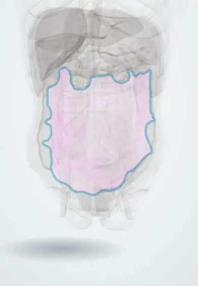
Peritoneal carcinomatosis is the spread of cancer over the peritoneal surfaces, most often from colorectal, gastric, ovarian, appendiceal or pancreatic cancer; rarely it starts in the peritoneum itself (peritoneal mesothelioma, primary peritoneal serous carcinoma). Pseudomyxoma peritonei is a special form in which a mucin-producing tumour, usually a ruptured low-grade appendiceal mucinous neoplasm, fills the abdomen with jelly-like mucin and slowly growing tumour cells.
Who gets it
Peritoneal spread is found in a meaningful share of colorectal and gastric cancers, at diagnosis or at recurrence, and in most women with advanced ovarian cancer. Pseudomyxoma peritonei is rare, about one to a few cases per million people a year, and commoner in women. In Colombia, gastric cancer is one of the most frequent causes of cancer death and is often diagnosed late, so carcinomatosis from stomach cancer is a common reason for malignant ascites in surgical wards.
Causes
- Colorectal cancer, especially mucinous, signet-ring or perforated tumours
- Gastric cancer, especially diffuse (signet-ring) type
- Ovarian, fallopian tube and primary peritoneal serous cancer
- Appendiceal mucinous neoplasms (the usual source of pseudomyxoma peritonei) and appendiceal adenocarcinoma
- Pancreatic and biliary cancer
- Primary peritoneal mesothelioma (asbestos exposure in some)
- Breast and lung cancer and melanoma (less often)
Risk factors
- Primary tumour that has grown through the serosa (T4)
- Tumour perforation or rupture, including at surgery
- Mucinous or signet-ring histology
- Ovarian or synchronous liver involvement
- Late diagnosis of gastric or ovarian cancer
Pathophysiology
The tumour grows through the outer wall of its organ (serosa) or ruptures → cells shed into the peritoneal cavity → they are carried by the peritoneal fluid current toward the absorbing surfaces (omental milky spots, diaphragmatic lymphatic openings) and the dependent pelvis → they attach, invade beneath the mesothelium and grow as nodules and plaques → the omentum thickens into an 'omental cake' → tumour blocks the lymphatic outflow and makes vessels leaky → malignant ascites → nodules tether and narrow bowel loops at several points → malignant bowel obstruction, malnutrition and cachexia. In pseudomyxoma peritonei, mucin-making cells escape from a ruptured appendiceal tumour → mucin collects where fluid is absorbed and pools (redistribution) → the abdomen slowly fills with jelly while the mobile small bowel stays relatively free.
Classification
- By origin: secondary (colorectal, gastric, ovarian, appendiceal, pancreatic) or primary peritoneal (mesothelioma, primary peritoneal serous carcinoma)
- Extent: Peritoneal Cancer Index (PCI), scoring lesion size 0–3 in 13 abdominal regions for a total of 0–39
- Completeness of cytoreduction score after surgery: CC-0 (no visible tumour) to CC-3 (large nodules left)
- Pseudomyxoma peritonei (PSOGI): acellular mucin, low-grade mucinous carcinoma peritonei, high-grade, and high-grade with signet-ring cells
- Ovarian cancer: peritoneal spread beyond the pelvis is FIGO stage III
Symptoms and signs
- Progressive abdominal swelling from ascites or mucin, with early fullness
- Vague abdominal pain and discomfort
- Nausea, vomiting and colicky pain from partial or complete bowel obstruction
- Loss of appetite, weight loss and muscle wasting despite a growing belly
- Shortness of breath from tense ascites or an associated pleural effusion
- Hard nodule at the umbilicus (Sister Mary Joseph nodule) or a rectal shelf on examination
- In women, an ovarian mass (mucinous appendiceal or gastric cancer can present as an ovarian tumour)
Diagnosis
- Clinical
- Suspect carcinomatosis in anyone with new ascites and no cirrhosis, weight loss, a known abdominal or ovarian cancer, or bowel obstruction without previous surgery. Examine the umbilicus, the rectum (rectal shelf), the ovaries and the left supraclavicular fossa.
- Tests
- Diagnostic paracentesis: serum–ascites albumin gradient usually below 1.1 g/dL with high protein, and cytology for malignant cells (positive in most cases of true carcinomatosis). Tumour markers to guide the search and follow-up: CEA, CA 19-9 and CA-125. Tissue diagnosis by image-guided biopsy or at laparoscopy; laparoscopy also measures the PCI before deciding on cytoreduction.
- Imaging
- CT of the chest, abdomen and pelvis with contrast: ascites, omental cake, nodular thickening of the peritoneum and mesentery, and the primary tumour; CT misses many nodules smaller than about 5 mm, so it underestimates extent. Pseudomyxoma: low-density material that scallops the surface of the liver and spleen. MRI with diffusion-weighted images and PET-CT in selected cases; ultrasound shows ascites and guides paracentesis.
Differential diagnosis
- Cirrhotic ascites
- Tuberculous peritonitis
- Heart failure or nephrotic syndrome ascites
- Spontaneous bacterial peritonitis
- Pancreatic ascites
- Benign mucinous cyst of the appendix or ovary without peritoneal spread
- Peritoneal lymphomatosis
New ascites: which cause?
| Condition | Typical patient | Ascitic fluid | Clinching finding |
|---|---|---|---|
| Peritoneal carcinomatosis | Known or hidden abdominal or ovarian cancer, weight loss | Low gradient (below 1.1), high protein, malignant cells | Positive cytology; omental cake and nodules on CT |
| Pseudomyxoma peritonei | Slowly enlarging belly, often a woman; appendiceal tumour | Thick jelly that will not flow through a needle | Scalloped liver surface on CT; mucin at surgery |
| Cirrhotic ascites | Alcohol or viral liver disease, stigmata of cirrhosis | High gradient (1.1 or more), low protein | Nodular liver and portal hypertension on ultrasound |
| Tuberculous peritonitis | Fever, night sweats; endemic area, HIV | Low gradient, lymphocytes, high adenosine deaminase | Caseating granulomas on peritoneal biopsy |
Goal of treatment
Find the primary cancer and the extent of spread, then choose between an attempt at cure (complete removal of all visible tumour, with or without heated intraperitoneal chemotherapy, in selected patients) and good palliation (relief of ascites and obstruction, nutrition and comfort).
Treatment
- Medical
- Systemic chemotherapy and targeted or immune therapy chosen by the primary cancer (colorectal, gastric, ovarian), alone in extensive disease or before and after surgery.
- Surgical
- Cytoreductive surgery in selected fit patients with limited, completely removable disease: peritonectomy of involved surfaces and resection of involved organs (often omentum, spleen, gallbladder, parts of bowel) with the aim of leaving no visible tumour (CC-0). Standard for pseudomyxoma peritonei and peritoneal mesothelioma, and an option for some colorectal and ovarian cancers; done in experienced centres.
- Procedure
- HIPEC (hyperthermic intraperitoneal chemotherapy) as a concept: after cytoreduction, heated chemotherapy solution is circulated through the abdomen during the operation to kill the microscopic cells left on the surfaces. Heat helps the drug penetrate and works against tumour cells; it only reaches a few millimetres, so it is useful only when the surgeon has removed all visible tumour. Its benefit is clearest in pseudomyxoma and ovarian cancer and debated in colorectal cancer.
- Procedure
- Palliation of ascites: therapeutic paracentesis when it causes symptoms, or a tunnelled indwelling peritoneal catheter for repeated drainage at home; diuretics help little in malignant ascites.
- Surgical
- Malignant bowel obstruction: nasogastric tube or venting gastrostomy, endoscopic stent for a single colonic or gastric outlet block, and bypass or stoma in selected patients; multiple levels of obstruction usually make surgery futile.
- Medical
- Symptom control for inoperable obstruction: antiemetics, opioid analgesia, antisecretory drugs (somatostatin analogues, anticholinergics) and corticosteroids, with early palliative care.
- Follow-up
- After cytoreduction: CT and tumour markers at intervals to detect recurrence, which is common and may be treated again in pseudomyxoma.
Complications
- Malignant bowel obstruction
- Tense ascites with breathlessness and pleural effusion
- Cachexia and malnutrition
- Ureteric obstruction and kidney failure
- Major morbidity after cytoreduction and HIPEC (anastomotic leak, fistula, bleeding, bone marrow suppression)
- Death from progressive disease
Common traps
- Ascites in a cancer patient is from liver metastases: carcinomatosis gives a low serum–ascites albumin gradient; a high gradient points to portal hypertension (cirrhosis or massive liver metastases)
- A normal CT rules out peritoneal spread: CT misses most nodules under about 5 mm; laparoscopy is how the extent is judged before a cure-intended operation
- HIPEC is the cure: the heated chemotherapy only penetrates millimetres; the surgery that removes all visible tumour does the main work, and HIPEC without complete cytoreduction is pointless
- Pseudomyxoma peritonei is an ovarian disease: in most cases the source is a ruptured appendiceal mucinous neoplasm, so the appendix is removed and examined even when an ovarian mass is found
- All carcinomatosis is incurable: selected patients with pseudomyxoma, mesothelioma and limited colorectal or ovarian spread live for years after complete cytoreduction
Prognosis
Carcinomatosis from gastric or pancreatic cancer usually means months of survival. Colorectal and ovarian carcinomatosis treated with modern chemotherapy, and complete cytoreduction in selected patients, can give years. Low-grade pseudomyxoma peritonei grows slowly; after complete cytoreduction with HIPEC most patients are alive many years later, while incomplete removal leads to repeated mucin build-up and obstruction.
Prevention
- Early diagnosis of gastric and colorectal cancer (screening, endoscopy for alarm symptoms)
- Avoid rupturing a tumour or mucinous appendix during surgery
- Proper staging, including diagnostic laparoscopy in gastric cancer, before major resections
Test yourself
Think of the answer, then tap the question to see it.
A 58-year-old without liver disease has new ascites; serum–ascites albumin gradient 0.8 g/dL with high protein. What is the most likely cause, and what test on the fluid comes next?
Peritoneal carcinomatosis (low-gradient ascites); send the fluid for cytology and search for the primary with CT and tumour markers.
Why does pseudomyxoma spare the small bowel surface while filling the omentum, the right subphrenic space and the pelvis?
Mucin moves with the peritoneal fluid current and collects where fluid is absorbed or pools (redistribution); the constantly moving small bowel sheds it, so it is involved late.
Why is HIPEC used only after the surgeon has removed all visible tumour?
Heated intraperitoneal chemotherapy only penetrates a few millimetres into tissue, so it can kill microscopic residual cells but not bulky nodules left behind.
A woman has a large mucinous ovarian mass and jelly in the abdomen at surgery. Which other organ must be removed and examined, and why?
The appendix: most pseudomyxoma peritonei comes from a ruptured appendiceal mucinous neoplasm, which often spreads to the ovaries and mimics an ovarian primary.
Why it happens
- Mucinous ovarian tumours
- Tumours of the appendix
- Metastases to the ovary
- Colorectal adenocarcinoma
- Diseases of the peritoneum
Connections
Educational information, not medical advice. For any symptom, see a health professional.
Sources: Pseudomyxoma Peritonei (StatPearls (NCBI Bookshelf), 2026); Cytoreduction (CRS) and Hyperthermic Intraperitoneal Chemotherapy (HIPEC) (StatPearls (NCBI Bookshelf), 2026); Peritoneal Cancer (StatPearls (NCBI Bookshelf), 2024); Ascites (StatPearls (NCBI Bookshelf), 2025).
Spotted a mistake?We are always improving for the community. Tell us what is wrong and our team will review it.Report a mistake
Representative model, for educational purposes only.