Conditions · Pediatrics and neonatology
Autosomal recessive polycystic kidney disease !
A rare inherited disease of babies with huge cystic kidneys, lung problems at birth from low amniotic fluid, and scarring of the liver.
Updated 2026-11-01
Structures involved
Tap one to see what it is and open it in 3D.
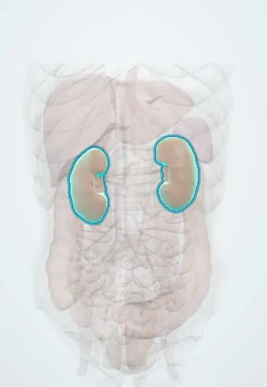
- Kidneys
- Liver
- Renal pelvis
- Portal vein
- Spleen
- Splenic vein
- Common hepatic duct
- Common bile duct
- Left renal artery
- Right renal artery
- Right lower lobe
- Left lower lobe
Why it happens
- Mendelian inheritance: autosomal dominant, recessive and X-linked
- Fibrosis in organs: TGF-β and the myofibroblast
- Cilia, microvilli and stereocilia
- Kidney: cortex, medulla and the nephron
What it is
An inherited ciliopathy (biallelic PKHD1 variants) in which the kidney collecting ducts dilate into tiny radial cysts and the bile ducts are malformed, causing congenital hepatic fibrosis. It usually starts before birth with huge kidneys, oligohydramnios and pulmonary hypoplasia, and survivors develop hypertension, chronic kidney disease and portal hypertension.
Who gets it
About 1 in 20,000 live births worldwide, with no sex predilection; carrier frequency about 1 in 70. Many cases are now detected on routine obstetric ultrasound. About 30% of affected newborns die in the first weeks, mainly from respiratory failure. No specific Colombian registry exists; affected children are managed in paediatric nephrology and transplant centres.
Causes
- Biallelic pathogenic variants in PKHD1 (encodes fibrocystin/polyductin, a protein of the primary cilium) in most cases
- Rarely variants in DZIP1L and other cilium genes
- Autosomal recessive: both parents are usually healthy carriers, with a 25% recurrence risk in each pregnancy
Risk factors
- Parents who are carriers or a previously affected child
- Parental consanguinity
- Two truncating PKHD1 variants are linked with the most severe perinatal form
Pathophysiology
Fibrocystin sits in the primary cilia of renal tubular and biliary epithelial cells. Its loss disturbs cell polarity and proliferation, so the collecting ducts dilate into fusiform cysts arranged radially from medulla to cortex; the kidneys keep their shape but become huge and echogenic. Poor fetal urine output causes oligohydramnios, which compresses the fetus and prevents normal lung growth (pulmonary hypoplasia), with flattened face, limb deformities and the Potter sequence. In the liver, failed remodelling of the ductal plate leaves dilated, abnormal bile ducts surrounded by portal fibrosis (congenital hepatic fibrosis), sometimes with dilated intrahepatic ducts (Caroli syndrome). Liver cell function is preserved, but fibrosis raises portal pressure and the abnormal ducts predispose to cholangitis.
Classification
- Perinatal: massive kidneys, oligohydramnios and pulmonary hypoplasia at birth
- Neonatal and infantile: kidney enlargement and hypertension in the first year
- Juvenile: milder kidney disease with predominant congenital hepatic fibrosis and portal hypertension in childhood or adolescence
Symptoms and signs
- Prenatal ultrasound with enlarged echogenic kidneys and oligohydramnios
- Newborn with respiratory distress, flattened face, clubfoot and other Potter features
- Large, firm, bilateral flank masses and a distended abdomen
- Early and often severe arterial hypertension
- Poor feeding, growth failure, polyuria from a urinary concentrating defect, and hyponatraemia in infancy
- Later: hepatosplenomegaly, low platelets, haematemesis from oesophageal varices, fever with jaundice (cholangitis)
Diagnosis
- Clinical
- Suspected from enlarged echogenic kidneys before or after birth, especially with oligohydramnios, hypertension or signs of liver fibrosis, when the parents have normal kidneys on ultrasound (which argues against the dominant form).
- Tests
- Serum creatinine, electrolytes (hyponatraemia), urinalysis and blood pressure; liver tests are usually near normal; full blood count for hypersplenism. Genetic testing for PKHD1 confirms the diagnosis and allows carrier testing and prenatal or preimplantation diagnosis in future pregnancies.
- Imaging
- Ultrasound is the key test: bilaterally enlarged, diffusely echogenic kidneys with loss of corticomedullary differentiation and many tiny cysts; increased liver echogenicity, splenomegaly and dilated bile ducts. Parents' kidney ultrasound helps exclude the dominant form. MRI or MR cholangiography and Doppler assess the liver, bile ducts and portal flow.
Differential diagnosis
- Autosomal dominant polycystic kidney disease with early onset
- Nephronophthisis and other ciliopathies (Bardet–Biedl, Meckel–Gruber syndromes)
- HNF1B-related kidney disease and multicystic dysplastic kidney
- Bilateral hydronephrosis from urinary obstruction
- Bilateral Wilms tumour or nephroblastomatosis; tuberous sclerosis with contiguous gene deletion
Goal of treatment
Support breathing at birth, control blood pressure, slow kidney decline and support growth, prepare for dialysis and transplantation, and prevent or treat the complications of portal hypertension and cholangitis.
Treatment
- Acute
- Neonatal intensive care: mechanical ventilation and surfactant or other respiratory support; nephrectomy of one or both kidneys may be needed if their size prevents breathing or feeding.
- Medical
- Antihypertensives, usually ACE inhibitors or angiotensin receptor blockers, often combined with other classes.
- Conservative
- Nutritional support (often by tube), sodium supplementation, correction of acidosis and anaemia, management of bone and mineral disorder; growth hormone may be used for poor growth.
- Procedure
- Peritoneal dialysis (preferred in infants) or haemodialysis for kidney failure; endoscopic band ligation for oesophageal varices.
- Surgical
- Kidney transplantation, or combined liver–kidney transplantation when liver disease is severe; portosystemic shunts in selected cases.
- Follow-up
- Lifelong follow-up: blood pressure, growth, renal function, platelet count and spleen size, and screening for varices; teach families the signs of cholangitis and bleeding; genetic counselling.
Complications
- Pulmonary hypoplasia, pneumothorax and neonatal death
- Severe hypertension with left ventricular hypertrophy
- Chronic kidney disease progressing to kidney failure in childhood or adolescence
- Portal hypertension with oesophageal varices, hypersplenism and bleeding
- Bacterial cholangitis and, rarely, cholangiocarcinoma
- Growth failure and developmental delay
Prognosis
The first weeks of life are decisive: about a third of newborns die, mostly of respiratory failure. Children who survive the newborn period have a good chance of reaching adulthood, but many need dialysis or a kidney transplant before adulthood and liver complications become the main problem later.
Prevention
- Genetic counselling for carrier parents (25% risk in each pregnancy); the disease itself cannot be prevented
- Prenatal or preimplantation genetic diagnosis when the family variants are known
Test yourself
Think of the answer, then tap the question to see it.
Which gene is mutated in most cases of ARPKD and what does its protein do?
PKHD1, encoding fibrocystin/polyductin, a protein of the primary cilium of tubular and biliary cells.
Which liver lesion is always present in ARPKD?
Congenital hepatic fibrosis from ductal plate malformation, sometimes with Caroli syndrome.
Why do newborns with ARPKD develop respiratory failure?
Low fetal urine output causes oligohydramnios, which leads to pulmonary hypoplasia (Potter sequence).
What is the typical ultrasound appearance of the kidneys?
Bilaterally enlarged, diffusely echogenic kidneys with many tiny cysts from dilated collecting ducts and poor corticomedullary differentiation.
How do you distinguish ARPKD from ADPKD in a child with cystic kidneys?
ARPKD is recessive, so parents usually have normal kidneys on ultrasound; it presents perinatally with hepatic fibrosis, whereas ADPKD usually has an affected parent and liver cysts rather than fibrosis.
Related conditions
- Autosomal dominant polycystic kidney disease
- Chronic kidney disease
- Arterial hypertension
- Portal hypertension
- Acute cholangitis
- Urinary tract infection (acute cystitis)
- Failure to thrive (faltering growth)
- Hyponatraemia
- Pneumothorax
- Neonatal respiratory distress syndrome (hyaline membrane disease)
- Tuberous sclerosis complex
- Wilms tumour (nephroblastoma)
- Anaemia of chronic disease (anaemia of inflammation)
Educational information, not medical advice. For any symptom, see a health professional.
Sources: Autosomal Recessive Polycystic Kidney Disease - NIDDK (National Institute of Diabetes and Digestive and Kidney Diseases (NIDDK), 2017); Polycystic kidney disease (MedlinePlus Genetics (U.S. National Library of Medicine), 2024).
Spotted a mistake?We are always improving for the community. Tell us what is wrong and our team will review it.Report a mistake
Representative model, for educational purposes only.