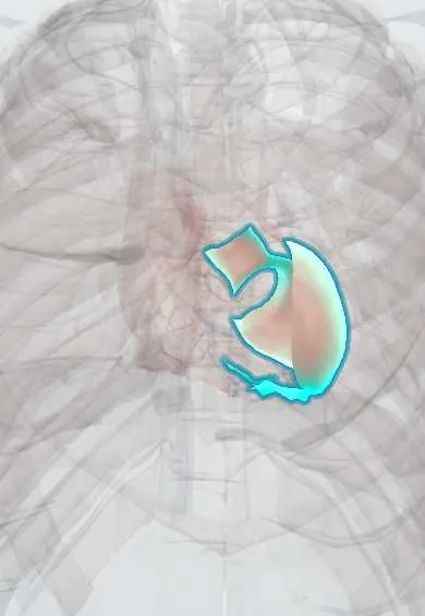
Cardiogenic shock !
Cardiogenic shock happens when the heart pumps so poorly that organs do not get enough blood. It is usually caused by a large heart attack.
Updated 2026-11-01
Structures involved
Tap one to see what it is and open it in 3D.
- Left ventricle
- LAD (anterior descending artery)
- Left ventricle papillary muscles
- Interventricular septum
- Right ventricle
- Right coronary artery
- Circumflex artery
In embryology
In the clinical exam
Why it happens
- Infarction: red vs white, and what decides the damage
- Shock: types, stages and the organs it damages
- The myocardial infarct over time: what the pathologist sees from hours to months
- Watershed areas and the organs most sensitive to ischemia
- Shock physiology
- The Frank–Starling mechanism
Show 1 more
What it is
A state of low cardiac output with tissue hypoperfusion caused by a primary heart problem, typically systolic blood pressure below about 90 mmHg with signs of poor organ perfusion despite adequate filling.
Who gets it
It complicates a minority of acute myocardial infarctions but is the leading cause of in-hospital death after infarction. In Colombia and Latin America, outcomes depend on timely access to primary angioplasty and intensive care, which is uneven outside large cities.
Causes
- Acute myocardial infarction with extensive left ventricular damage (the most common cause)
- Mechanical complications of infarction: papillary muscle rupture with acute mitral regurgitation, ventricular septal rupture, free wall rupture
- Right ventricular infarction
- End-stage cardiomyopathy or decompensated heart failure, acute myocarditis
- Severe valve disease and sustained arrhythmias
Risk factors
- Large anterior infarction (proximal LAD occlusion) or multivessel disease
- Older age, diabetes and previous infarction or heart failure
- Delayed reperfusion
Pathophysiology
Loss of contractile myocardium lowers stroke volume and cardiac output. Low blood pressure reduces coronary perfusion, which causes more ischaemia and a downward spiral. Compensatory vasoconstriction and fluid retention raise filling pressures and cause pulmonary congestion, and the inflammatory response can add vasodilatation. The classic profile is «cold and wet»: high wedge pressure, low cardiac index and high systemic resistance.
Classification
- Haemodynamic profile: cold and wet (most common), cold and dry
- Severity staging from at risk to refractory shock (SCAI stages A to E)
- Predominantly left, right or biventricular failure
Symptoms and signs
- Hypotension with a weak, fast pulse
- Cold, clammy, mottled skin and delayed capillary refill
- Confusion or agitation
- Low urine output
- Dyspnoea and lung crackles from pulmonary oedema
- Chest pain when the cause is an infarction
Diagnosis
- Clinical
- Hypotension with signs of hypoperfusion (cold skin, confusion, oliguria) and usually congestion; examination for new murmurs, jugular venous pressure and lung crackles.
- Tests
- 12-lead ECG (ST elevation, right-sided leads), troponin, lactate, arterial blood gas, kidney and liver function, natriuretic peptides; invasive haemodynamics (pulmonary artery catheter) in selected cases: low cardiac index and high wedge pressure.
- Imaging
- Urgent echocardiography to assess ventricular function and exclude mechanical complications and tamponade; coronary angiography to find the culprit lesion; chest X-ray for pulmonary oedema.
Differential diagnosis
- Hypovolaemic shock
- Septic (distributive) shock
- Obstructive shock: massive pulmonary embolism, cardiac tamponade, tension pneumothorax
- Anaphylaxis
Goal of treatment
Restore perfusion quickly by treating the cause (above all, opening the infarct-related artery), supporting blood pressure and cardiac output, and preventing multi-organ failure.
Treatment
- Acute
- Oxygen or ventilatory support as needed, arterial line, careful fluids only if there is no congestion (for example in right ventricular infarction).
- Procedure
- Emergency coronary revascularisation, usually percutaneous coronary intervention of the culprit artery; surgery for mechanical complications or anatomy unsuitable for stenting.
- Medical
- Vasopressors (norepinephrine is preferred) to keep perfusion pressure and inotropes (dobutamine, milrinone) to raise cardiac output; loop diuretics for congestion once pressure allows.
- Procedure
- Temporary mechanical circulatory support in selected patients: intra-aortic balloon pump, percutaneous ventricular assist devices or extracorporeal membrane oxygenation (ECMO), as a bridge to recovery, transplant or a durable device.
- Follow-up
- Intensive care monitoring: blood pressure, heart rhythm, hourly urine output, lactate trend, mental status and kidney function; nursing care focused on perfusion and safety.
Complications
- Multi-organ failure: acute kidney injury, ischaemic liver injury, bowel ischaemia
- Ventricular arrhythmias and cardiac arrest
- Respiratory failure
- Bleeding and limb ischaemia from devices and arterial access
- Death
Prognosis
Mortality remains high, around half of patients in many series. Early revascularisation of the culprit artery is the intervention that has most clearly improved survival.
Prevention
- Fast recognition and reperfusion of myocardial infarction
- Optimal treatment of heart failure
Test yourself
Think of the answer, then tap the question to see it.
What is the most common cause of cardiogenic shock?
Acute myocardial infarction with extensive left ventricular damage.
What is the classic haemodynamic profile of cardiogenic shock?
Low cardiac index, high pulmonary capillary wedge pressure and high systemic vascular resistance: «cold and wet».
Which treatment most clearly lowers mortality in shock due to infarction?
Early revascularisation of the infarct-related artery, usually by percutaneous coronary intervention.
Which vasopressor is generally preferred in cardiogenic shock?
Norepinephrine, often combined with an inotrope such as dobutamine.
A patient with an inferior infarction is hypotensive with clear lungs and distended neck veins. What is suspected?
Right ventricular infarction; it is preload-dependent, so nitrates are avoided and cautious fluids are given.
Related conditions
- Myocardial Infarction (Heart Attack)
- Heart failure
- Mitral regurgitation
- Ventricular tachycardia
- Cardiac arrest (VF, pulseless VT, asystole, PEA)
- Cardiac tamponade
- Pulmonary embolism
- Hypovolaemic shock
- Sepsis and septic shock
Educational information, not medical advice. For any symptom, see a health professional.
Sources: Cardiogenic shock: MedlinePlus Medical Encyclopedia (MedlinePlus, US National Library of Medicine, 2026).
Spotted a mistake?We are always improving for the community. Tell us what is wrong and our team will review it.Report a mistake
Representative model, for educational purposes only.