Conditions · Pediatrics and neonatology
Common variable immunodeficiency (CVID) !
An immune disorder where the body makes too few antibodies, causing repeated lung, sinus and gut infections, often found in young adults.
Updated 2026-11-01
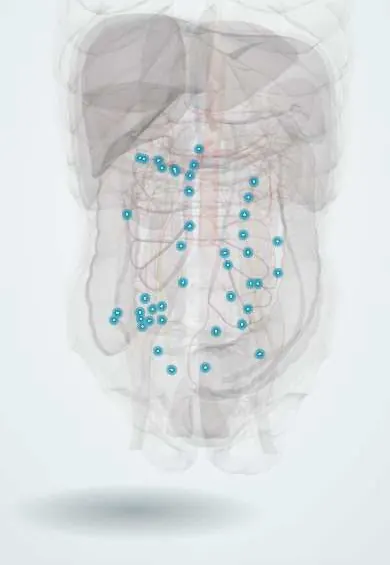
Structures involved
Tap one to see what it is and open it in 3D.
Why it happens
What it is
A primary antibody deficiency in which B cells fail to mature into antibody-producing plasma cells. Serum IgG is markedly low, with low IgA and/or IgM, and the response to vaccines is poor. It is the most common symptomatic primary immunodeficiency in adults and also causes autoimmunity, granulomatous disease, lymphoid hyperplasia and cancer.
Who gets it
Estimated at about 1 in 25,000 to 50,000 people, affecting men and women equally. Onset can be at any age, but most patients are diagnosed in their twenties or thirties, often after years of delay. In Latin America, registries of the Latin American Society for Immunodeficiencies (LASID) show CVID as one of the most frequently reported antibody deficiencies, and under-diagnosis is common.
Causes
- Usually no single cause is found; most cases are sporadic and probably polygenic
- A monogenic cause is identified in a minority: variants in TNFRSF13B (TACI, the most frequent), NFKB1, NFKB2, ICOS, CD19, LRBA, CTLA4 and others
- Exclusion of secondary hypogammaglobulinaemia is required (protein loss, lymphoma or myeloma, drugs such as anticonvulsants or B-cell-depleting therapy)
Risk factors
- Family history of CVID or selective IgA deficiency (both can occur in the same family)
- Recurrent sinopulmonary bacterial infections since adolescence or early adulthood
Pathophysiology
Defective B-cell activation, class switching and terminal differentiation lead to few switched memory B cells and plasma cells. Without enough IgG and secretory IgA, encapsulated bacteria (Streptococcus pneumoniae, Haemophilus influenzae) repeatedly infect the airways, and Giardia and other pathogens infect the gut. Immune dysregulation coexists, so patients develop autoimmune cytopenias, non-caseating granulomas, lymphoid hyperplasia of the gut and spleen, and lymphoma.
Classification
- Infection-only phenotype: recurrent infections without other complications
- Complicated (non-infectious) phenotypes: autoimmunity, granulomatous-lymphocytic interstitial lung disease, enteropathy, lymphoproliferation, lymphoma
Symptoms and signs
- Recurrent bacterial sinusitis, otitis, bronchitis and pneumonia
- Chronic productive cough from bronchiectasis
- Chronic diarrhoea, malabsorption and weight loss (Giardia, celiac-like enteropathy)
- Lymphadenopathy and splenomegaly
- Autoimmune features: bruising or bleeding from low platelets, anaemia from haemolysis, arthritis
- Cough and breathlessness from granulomatous interstitial lung disease
Diagnosis
- Clinical
- Suspect it in an adolescent or adult with recurrent sinopulmonary infections, bronchiectasis, chronic diarrhoea or unexplained autoimmune cytopenia. Diagnosis needs low IgG plus low IgA and/or IgM, poor vaccine responses and exclusion of secondary causes; typically age over 4 years.
- Tests
- Serum IgG, IgA and IgM; specific antibody response to protein and polysaccharide vaccines (tetanus, pneumococcal); B-cell and switched memory B-cell counts by flow cytometry; complete blood count; stool tests for Giardia; serum protein electrophoresis to exclude protein loss or myeloma; genetic panel in selected cases. Celiac serology is unreliable because antibody production is impaired.
- Imaging
- Chest high-resolution CT for bronchiectasis and granulomatous-lymphocytic interstitial lung disease; abdominal imaging for splenomegaly and lymphadenopathy; biopsy of enlarging nodes.
Differential diagnosis
- X-linked agammaglobulinaemia (boys, onset in infancy, absent B cells)
- Selective IgA deficiency
- Secondary hypogammaglobulinaemia (protein-losing enteropathy, nephrotic syndrome, lymphoma, myeloma, drugs)
- Cystic fibrosis and primary ciliary dyskinesia
- Sarcoidosis (for granulomatous disease)
Goal of treatment
Prevent infections and lung damage with lifelong immunoglobulin replacement, treat infections early, and detect and manage autoimmune, granulomatous and malignant complications.
Treatment
- Medical
- Lifelong immunoglobulin (IgG) replacement, intravenous or subcutaneous, adjusted to keep trough levels and infections under control.
- Medical
- Prompt antibiotics for bacterial infections; prophylactic antibiotics in some patients with bronchiectasis.
- Medical
- Treatment of Giardia and other gut infections with antiparasitic agents.
- Medical
- Immunosuppressive therapy (corticosteroids, rituximab, others) for autoimmune cytopenias, granulomatous lung disease or enteropathy.
- Follow-up
- Monitoring: lung function and chest CT, blood counts, liver tests and surveillance for lymphoma and gastric cancer.
- Conservative
- Airway clearance physiotherapy; live vaccines are avoided, and inactivated vaccines are not relied on for protection.
Complications
- Bronchiectasis and chronic lung disease
- Autoimmune cytopenias (immune thrombocytopenia, autoimmune haemolytic anaemia)
- Granulomatous-lymphocytic interstitial lung disease and liver granulomas
- Enteropathy with malabsorption, and nodular lymphoid hyperplasia of the bowel
- Non-Hodgkin lymphoma and gastric cancer
- Infusion reactions to immunoglobulin
Prognosis
With regular immunoglobulin replacement, patients who have only infections have a near-normal life expectancy. Survival is lower in those with non-infectious complications such as chronic lung disease, liver disease or lymphoma.
Prevention
- No primary prevention is known
- Early diagnosis and immunoglobulin replacement prevent bronchiectasis
- Vaccination of household contacts
Test yourself
Think of the answer, then tap the question to see it.
What laboratory profile defines CVID?
Low IgG with low IgA and/or IgM, poor vaccine antibody responses and no secondary cause.
Which gut parasite commonly causes chronic diarrhoea in CVID?
Giardia lamblia (giardiasis).
What is the mainstay of treatment?
Lifelong immunoglobulin (IgG) replacement therapy, intravenous or subcutaneous.
Which two malignancies have an increased risk in CVID?
Non-Hodgkin lymphoma and gastric cancer.
How is CVID distinguished from X-linked agammaglobulinaemia?
CVID usually starts later and B cells are present (though dysfunctional); XLA starts in infant boys and B cells are absent.
Related conditions
- Bronchiectasis
- Giardiasis
- Non-Hodgkin lymphoma
- Immune thrombocytopenia (ITP)
- Autoimmune haemolytic anaemia
- Gastric cancer
- X-linked (Bruton) agammaglobulinaemia
- Selective IgA deficiency
- Sarcoidosis
- Community-acquired pneumonia
- Chronic rhinosinusitis and nasal polyps
Educational information, not medical advice. For any symptom, see a health professional.
Sources: Common variable immune deficiency (MedlinePlus Genetics (U.S. National Library of Medicine), 2016).
Spotted a mistake?We are always improving for the community. Tell us what is wrong and our team will review it.Report a mistake
Representative model, for educational purposes only.