
Craniosynostosis: orbital and eye features
Early fusion of skull sutures, as in Crouzon or Apert syndrome, gives shallow orbits and bulging eyes, risking exposure, raised pressure and squint.
What it is
Craniosynostosis is premature fusion of skull sutures. Syndromic forms such as Crouzon and Apert (FGFR2) give shallow orbits with proptosis, wide-set eyes and midface hypoplasia, with risks of corneal exposure, papilloedema and optic atrophy from raised pressure, V-pattern strabismus, refractive error and amblyopia.
Who gets it
Craniosynostosis affects about 1 in 2,000-2,500 births; syndromic forms are a minority. Crouzon and Apert syndromes are rare and usually autosomal dominant, often new mutations.
Causes
- Non-syndromic single-suture synostosis (sagittal most common)
- Crouzon syndrome: FGFR2 mutation (with acanthosis nigricans in FGFR3 variant)
- Apert syndrome: FGFR2 mutation with syndactyly of hands and feet
- Pfeiffer, Muenke and Saethre-Chotzen syndromes (FGFR1/2/3, TWIST1)
Risk factors
- Family history
- Advanced paternal age (new FGFR2 mutations)
Pathophysiology
Premature fusion stops growth across the suture, with compensatory growth elsewhere. In coronal and syndromic synostosis the orbital roof and midface are retruded, creating shallow orbits and proptosis. Restricted skull growth can raise intracranial pressure, causing papilloedema and later optic atrophy. Excyclorotation of the orbits and abnormal muscle insertions cause V-pattern strabismus and apparent inferior oblique overaction.
Classification
- Non-syndromic: sagittal, metopic, unicoronal, lambdoid
- Syndromic: Crouzon, Apert, Pfeiffer, Muenke, Saethre-Chotzen
Symptoms and signs
- Abnormal head shape
- Bulging eyes, wide-set eyes, eyelids that may not close
- Squint, especially exotropia with V pattern
- Headache, vomiting or developmental delay (raised pressure)
- Breathing problems in sleep (midface hypoplasia)
Diagnosis
- Clinical
- Examination of skull shape, sutures, face and limbs; eye examination for exposure, proptosis, strabismus, refraction, amblyopia and optic discs in every syndromic child.
- Tests
- Genetic testing for FGFR and TWIST1 mutations; visual evoked potentials or optical coherence tomography of the disc in young children; sleep studies.
- Imaging
- Low-dose CT with 3D reconstruction of the skull to confirm suture fusion and orbital shape; MRI for hydrocephalus or Chiari malformation.
Differential diagnosis
- Positional (deformational) plagiocephaly
- Microcephaly
- Thyroid eye disease (proptosis in adults)
- Orbital tumours
Unusual head shape or bulging eyes in a child: which one?
| Condition | Sutures | Eyes and orbits | Key clue |
|---|---|---|---|
| Syndromic craniosynostosis (Crouzon, Apert) | Several fused early, often both coronal | Shallow orbits, proptosis, wide-set eyes, V-pattern strabismus | Midface hypoplasia; syndactyly in Apert; FGFR2 mutation |
| Non-syndromic single-suture synostosis | One fused (sagittal most common) | Usually normal; unicoronal synostosis can tilt one orbit | Head shape set by which suture fused |
| Positional plagiocephaly | Open | Normal | Flat area from lying position; improves with repositioning |
| Thyroid eye disease | Open | Proptosis from swollen muscles and fat; lid retraction | Adults with thyroid disease; not present from birth |
Goal of treatment
Allow normal brain growth, protect the eyes and vision and improve function and appearance; care is in a craniofacial team.
Treatment
- Surgical
- Cranial vault remodelling and fronto-orbital advancement in infancy; midface (Le Fort III or monobloc) advancement to deepen the orbits.
- Conservative
- Lubricants and moisture chambers for exposure; glasses and patching for refractive error and amblyopia.
- Surgical
- Tarsorrhaphy for severe exposure; strabismus surgery for V pattern and other deviations.
- Follow-up
- Regular fundus and vision checks for papilloedema and optic atrophy throughout childhood.
Complications
- Exposure keratopathy and corneal scarring
- Optic atrophy from chronic raised pressure
- Amblyopia
- Globe luxation
Common traps
- Proptosis in Crouzon syndrome means the eyes are too big: the eyes are normal in size but the orbits are too shallow, which is why midface and orbital advancement lets the lids close again
- Normal vision at one visit means raised pressure is not a worry: papilloedema can be silent in young children and optic atrophy is the main cause of permanent loss, so fundus checks continue throughout childhood
- A flat back of the head in an infant is usually craniosynostosis: positional plagiocephaly from lying position is far more common and leaves the sutures open; true lambdoid synostosis is rare
- Apert and Crouzon syndromes are told apart by their gene: both are usually FGFR2 mutations; Apert is recognised clinically by syndactyly of the hands and feet
- The V pattern always means true inferior oblique overaction: in craniosynostosis it is often apparent, from excyclorotated orbits and displaced muscle insertions, so surgery is planned around the orbital anatomy
Prognosis
With early surgery and eye follow-up, most children keep good vision; vision loss usually comes from untreated raised pressure or exposure.
Test yourself
Think of the answer, then tap the question to see it.
Why do children with Crouzon syndrome have bulging eyes although the globes are normal in size?
The orbital roof and midface fail to grow forward, so the orbits are too shallow for normal eyes, which are pushed forward and may not be covered by the lids.
A child with Apert syndrome develops headaches and swollen optic discs. What is the concern, and why does it matter for vision?
Raised intracranial pressure from a skull too small for the growing brain. Untreated, papilloedema progresses to optic atrophy, the main cause of permanent vision loss in these children.
An infant with Crouzon syndrome has one eye pushed in front of the lids after crying. What is this, and what is the immediate aim?
Globe luxation: the shallow orbit lets the eye slip past the lids. The aim is to reposition the eye behind the lids promptly and protect the cornea; tarsorrhaphy or orbital advancement may follow.
Which feature distinguishes Apert from Crouzon syndrome, and what do they share?
Apert has syndactyly of the hands and feet. Both usually carry FGFR2 mutations and share shallow orbits with proptosis and midface hypoplasia.
Why is V-pattern strabismus common in syndromic craniosynostosis?
The orbits are excyclorotated, so muscle insertions and pulleys sit in abnormal positions; the inferior obliques appear to overact and the eyes diverge in upgaze.
In pediatrics
Connections
Educational information, not medical advice. For any symptom, see a health professional.
Sources: Craniosynostosis (StatPearls Publishing (NCBI Bookshelf), 2025); Crouzon Syndrome (StatPearls Publishing (NCBI Bookshelf), 2025); Apert Syndrome (StatPearls Publishing (NCBI Bookshelf), 2025).
Spotted a mistake?We are always improving for the community. Tell us what is wrong and our team will review it.Report a mistake
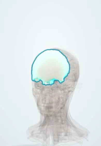
Representative model, for educational purposes only.