Conditions · Infectious and tropical diseases
Human African trypanosomiasis (sleeping sickness) !
A parasite spread by the tsetse fly in rural Africa that first causes fever and swollen neck nodes, then invades the brain and disturbs sleep.
Updated 2026-11-01
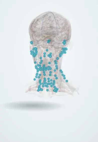
Structures involved
Tap one to see what it is and open it in 3D.
Microorganisms that cause it
Why it happens
- Wakefulness, sleep, the EEG and seizures
- Functions of the hypothalamus
- CSF and the blood–brain barrier
- Immunoglobulins (antibodies)
What it is
Infection by Trypanosoma brucei, transmitted by the tsetse fly in sub-Saharan Africa. It has a first blood-and-lymph stage and a second stage in which the parasite crosses into the central nervous system, causing meningoencephalitis with a disturbed sleep-wake cycle. Without treatment it is usually fatal. It does not occur in the Americas, unlike Chagas disease (T. cruzi).
Who gets it
Endemic only in sub-Saharan Africa, in rural people who farm, fish, keep animals or hunt. T. b. gambiense (West and Central Africa) causes about 92% of cases and T. b. rhodesiense (East and Southern Africa) about 8%. Cases fell by 97% in 20 years to fewer than 1,000 a year, most in the Democratic Republic of the Congo. In Colombia it is a travel or migration diagnosis; local T. cruzi and the harmless T. rangeli must not be confused with it.
Causes
- Trypanosoma brucei gambiense: chronic form; humans are the main reservoir
- Trypanosoma brucei rhodesiense: acute form; wild and domestic animals, especially cattle, are reservoirs
- Vector: tsetse fly (Glossina) bite
- Rarely: mother to baby, blood transfusion, transplant, sexual contact or laboratory accidents
Risk factors
- Rural life or work in endemic areas (farming, fishing, hunting, herding)
- Visits to game parks and safaris in East Africa (rhodesiense)
- Living near rivers and forests where tsetse flies breed
- Weak surveillance and access to health care in conflict areas
Pathophysiology
The tsetse bite injects trypomastigotes that multiply locally (chancre), then in blood and lymph. The parasite evades antibodies by constantly switching its variant surface glycoprotein, producing waves of parasitaemia and fever and very high polyclonal IgM. In the second stage it crosses the blood-brain barrier and causes perivascular lymphocytic infiltrates (perivascular cuffing), with morular (Mott) plasma cells; inflammation of the hypothalamus and brainstem disrupts circadian rhythm and sleep.
Classification
- By subspecies: gambiense (chronic, months to years) and rhodesiense (acute, weeks to months)
- Stage 1: haemolymphatic
- Stage 2: meningoencephalitic, defined by parasites or raised white cells in cerebrospinal fluid
Symptoms and signs
- Painful nodule or chancre at the bite site (more common in rhodesiense and in travellers)
- Intermittent fever, headache, joint pain and itching
- Enlarged, soft posterior cervical lymph nodes (Winterbottom sign) in gambiense disease
- Later: daytime sleepiness and nighttime insomnia, behaviour and personality changes, confusion
- Tremor, poor coordination, painful sensitivity to touch and, finally, coma
Diagnosis
- Clinical
- Suspected from exposure history, fever and lymphadenopathy, and later from sleep and behaviour changes; travellers usually have rhodesiense disease with a chancre.
- Tests
- Screening with the card agglutination test (CATT) or rapid serological tests for gambiense; confirmation by finding trypanosomes in blood, lymph node aspirate or chancre fluid (thick smear and concentration methods). Staging by lumbar puncture: parasites or more than 5 white cells per microlitre in cerebrospinal fluid define stage 2. High IgM supports the diagnosis.
- Imaging
- Not needed for diagnosis; brain MRI may show white matter and basal ganglia changes in advanced disease.
Differential diagnosis
- Malaria
- HIV infection and tuberculosis (lymphadenopathy and fever)
- Typhoid fever
- Bacterial meningitis and viral encephalitis
- Psychiatric illness, especially in stage 2
Goal of treatment
Diagnose early, choose drugs by subspecies and stage, cure the infection with the least toxic option and confirm the absence of relapse.
Treatment
- Medical
- Gambiense disease: oral fexinidazole for stage 1 and non-severe stage 2; pentamidine for stage 1; nifurtimox-eflornithine combination (NECT) for severe stage 2.
- Medical
- Rhodesiense disease: suramin for stage 1 and melarsoprol for stage 2, an arsenical that can cause a fatal reactive encephalopathy; newer oral drugs are expanding options.
- Follow-up
- Hospital monitoring during treatment, especially for drug toxicity, and follow-up with lumbar puncture as indicated (traditionally every six months for two years).
Complications
- Coma and death if untreated
- Permanent neurological and psychiatric sequelae
- Myocarditis and heart failure (especially rhodesiense)
- Severe anaemia and endocrine disorders
- Treatment-related encephalopathy with melarsoprol
Prognosis
Untreated, it is almost always fatal: rhodesiense within weeks to months and gambiense within months to years. Early treatment cures most patients; stage 2 disease leaves more sequelae.
Prevention
- Active screening and treatment of populations at risk (gambiense)
- Tsetse fly control with traps and insecticide-treated targets
- Neutral-coloured long clothing, insect repellent and avoiding bushes; there is no vaccine or chemoprophylaxis
- Treating cattle reservoirs (rhodesiense)
Test yourself
Think of the answer, then tap the question to see it.
Which vector transmits African trypanosomiasis?
The tsetse fly (Glossina).
What is Winterbottom sign?
Enlarged posterior cervical lymph nodes, typical of gambiense disease.
How do trypanosomes evade the immune response?
By antigenic variation of their variant surface glycoprotein, producing waves of parasitaemia.
Which test defines stage 2 disease?
Lumbar puncture showing parasites or raised white cells in the cerebrospinal fluid.
How does African trypanosomiasis differ geographically from Chagas disease?
It occurs only in sub-Saharan Africa (T. brucei, tsetse fly), while Chagas is American (T. cruzi, triatomine bugs).
Related conditions
- Chagas disease
- Malaria
- HIV infection and AIDS
- Bacterial meningitis
- Viral encephalitis (herpes simplex encephalitis)
- Cervical lymphadenopathy
- Tuberculosis
- Typhoid (enteric) fever
Educational information, not medical advice. For any symptom, see a health professional.
Sources: Trypanosomiasis, human African (sleeping sickness) (World Health Organization, 2023); About Sleeping Sickness | Sleeping Sickness (African Trypanosomiasis) | CDC (Centers for Disease Control and Prevention, 2024).
Spotted a mistake?We are always improving for the community. Tell us what is wrong and our team will review it.Report a mistake
Representative model, for educational purposes only.