Conditions · Hematology and oncology
Hypercalcaemia of malignancy !
Some cancers raise blood calcium by releasing a hormone-like protein or by destroying bone, causing thirst, confusion, constipation and kidney injury.
Updated 2026-11-01
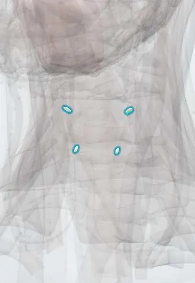
Structures involved
Tap one to see what it is and open it in 3D.
Why it happens
- Paraneoplastic syndromes: the table to know
- Pathologic calcification: dystrophic vs metastatic
- Pathways of spread and the usual metastatic sites
- Calcium regulation: PTH, vitamin D and calcitonin
- Bone remodeling
- Ossification, growth plate and bone remodeling
What it is
A high blood calcium caused by cancer. It is the most frequent life-threatening metabolic complication of malignancy and usually appears in patients with advanced, known disease. Unlike primary hyperparathyroidism, which is mild and chronic, it tends to rise quickly, cause symptoms, and come with a suppressed parathyroid hormone (PTH).
Who gets it
Affects up to about a fifth of patients with cancer at some point in their illness. The most commonly associated tumours are multiple myeloma, breast cancer, squamous cell lung cancer, head and neck and kidney cancers, and some lymphomas (including adult T-cell leukaemia/lymphoma linked to HTLV-1, which is endemic in parts of Colombia's Pacific coast and the Caribbean). Its appearance usually marks advanced disease.
Causes
- Humoral hypercalcaemia (most cases): tumour secretes PTH-related protein (PTHrP), typical of squamous cell carcinomas, renal and breast cancers
- Local osteolytic hypercalcaemia: bone metastases or myeloma cells activate osteoclasts (breast cancer, multiple myeloma)
- Excess active vitamin D (calcitriol) made by lymphomas
- Rare: true ectopic PTH secretion; parathyroid carcinoma
Risk factors
- Multiple myeloma and extensive bone metastases
- Squamous cell carcinomas (lung, head and neck, oesophagus, cervix)
- Breast, kidney and ovarian cancer; lymphoma
- Dehydration, immobility and kidney impairment
- Thiazide diuretics or calcium and vitamin D supplements in a patient with cancer
Pathophysiology
PTHrP binds the PTH receptor, stimulating osteoclast bone resorption and calcium reabsorption in the kidney. In osteolytic disease, tumour cells release cytokines and RANKL that activate osteoclasts locally. High calcium impairs the kidney's ability to concentrate urine (nephrogenic diabetes insipidus), so the patient loses salt and water; the resulting volume depletion lowers glomerular filtration and calcium excretion, a vicious circle that drives calcium higher. Raised calcium also depresses nerve and muscle excitability and shortens the QT interval.
Classification
- By mechanism: humoral (PTHrP), local osteolytic, calcitriol-mediated, ectopic PTH
- By severity of corrected total calcium: mild, moderate and severe (severe roughly above 14 mg/dL, or 3.5 mmol/L)
Symptoms and signs
- Thirst and passing large amounts of urine, with dehydration
- Nausea, vomiting, loss of appetite and constipation
- Fatigue, muscle weakness and bone pain
- Confusion, drowsiness, depression, progressing to stupor or coma
- Abdominal pain (peptic ulcer, pancreatitis) and kidney stones in longer-standing cases
Diagnosis
- Clinical
- Suspect it in any patient with cancer who becomes confused, dehydrated, constipated or vomits. Assess volume status, mental state and medications (thiazides, calcium, vitamin D, lithium).
- Tests
- Total calcium corrected for albumin, or ionised calcium. Intact PTH is the key first step: it is low or suppressed in malignancy and high in primary hyperparathyroidism. PTHrP, 25-hydroxy and 1,25-dihydroxy vitamin D help define the mechanism. Kidney function, electrolytes, magnesium and phosphate; serum protein electrophoresis and free light chains if myeloma is possible. ECG for short QT and arrhythmias.
- Imaging
- Imaging searches for the cancer and bone lesions: chest X-ray or CT, skeletal survey or whole-body imaging for myeloma, bone scan for metastases (not useful for myeloma lesions).
Differential diagnosis
- Primary hyperparathyroidism (high or inappropriately normal PTH; can coexist with cancer)
- Granulomatous disease (sarcoidosis, tuberculosis) with excess calcitriol
- Vitamin D or calcium intoxication, thiazide or lithium effect
- Immobilisation, thyrotoxicosis, adrenal insufficiency
- Familial hypocalciuric hypercalcaemia
Goal of treatment
Restore fluid volume, lower calcium safely and quickly, protect the kidneys and brain, and control the underlying cancer, which is what keeps calcium down.
Treatment
- Acute
- Intravenous isotonic saline is the first measure, to correct dehydration and increase calcium excretion; loop diuretics are used only once the patient is rehydrated or if fluid overload appears.
- Medical
- Calcitonin acts within hours but its effect wears off in about two days; it bridges the gap until slower drugs work.
- Medical
- Intravenous bisphosphonates are the main drug class: they block osteoclasts, lower calcium over 2 to 4 days and need kidney-function adjustment; denosumab (an anti-RANKL antibody) is used when bisphosphonates fail or kidneys are impaired.
- Medical
- Corticosteroids for calcitriol-mediated hypercalcaemia (lymphoma) and steroid-sensitive tumours; stop thiazides, calcium and vitamin D supplements.
- Procedure
- Haemodialysis with a low-calcium bath for severe, refractory hypercalcaemia or when kidney or heart failure prevents fluid therapy.
- Follow-up
- Nursing priorities: strict fluid balance and daily weight, watch for fluid overload, frequent calcium and electrolyte checks, cardiac monitoring, neurological status and fall prevention, and encourage mobility.
Complications
- Acute kidney injury, nephrocalcinosis and kidney stones
- Arrhythmias and cardiac arrest
- Coma and death
- Pancreatitis and peptic ulcer
- Hypocalcaemia or osteonecrosis of the jaw from anti-resorptive treatment
Prognosis
Calcium usually responds to fluids and anti-resorptive drugs within days, but the condition signals advanced cancer: without effective cancer treatment, many patients survive only a few months. Outlook is better in myeloma, lymphoma and breast cancer when the tumour responds to therapy.
Prevention
- Periodic calcium checks in patients with myeloma or bone metastases
- Bone-protective drugs in patients with bone metastases, as part of oncology care
- Avoid dehydration, prolonged immobility and thiazide diuretics in high-risk patients
Test yourself
Think of the answer, then tap the question to see it.
What is the most common mechanism of hypercalcaemia of malignancy?
Tumour secretion of PTH-related protein (humoral hypercalcaemia), typical of squamous cell carcinomas.
Which laboratory finding separates hypercalcaemia of malignancy from primary hyperparathyroidism?
Intact PTH: suppressed in malignancy, high or inappropriately normal in primary hyperparathyroidism.
What is the first treatment for symptomatic hypercalcaemia of malignancy?
Intravenous isotonic saline to restore volume and increase urinary calcium excretion.
Which drug class is the mainstay for lowering calcium over the following days?
Intravenous bisphosphonates, which inhibit osteoclast bone resorption.
Which ECG change is classic in severe hypercalcaemia?
A shortened QT interval, with risk of arrhythmias.
Related conditions
- Hypercalcaemia
- Primary hyperparathyroidism
- Multiple myeloma
- Lung cancer
- Breast cancer
- Renal cell carcinoma
- Non-Hodgkin lymphoma
- Acute kidney injury
- Kidney stones (urolithiasis)
- Dehydration (fluid volume deficit)
- Sarcoidosis
- Acute pancreatitis
- Constipation and faecal impaction
- Malignant spinal cord compression
- Tumour lysis syndrome
Educational information, not medical advice. For any symptom, see a health professional.
Sources: Hypercalcemia: MedlinePlus Medical Encyclopedia (MedlinePlus (U.S. National Library of Medicine), 2026); Cancer-related hypercalcemia and potential treatments (Frontiers in Endocrinology (PubMed Central), 2023).
Spotted a mistake?We are always improving for the community. Tell us what is wrong and our team will review it.Report a mistake
Representative model, for educational purposes only.