Conditions · Hematology and oncology
Malignant spinal cord compression !
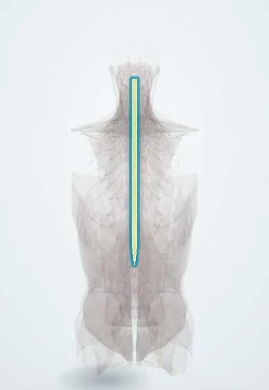
Cancer that has spread to the spine presses on the spinal cord. It starts with back pain and can cause paralysis unless treated within hours.
Updated 2026-11-01
Structures involved
Tap one to see what it is and open it in 3D.
- Spinal cord
- T6 vertebra
- L1 vertebra
- Dura (dural sac)
- Cauda equina
- Mid-back nerve roots
- T4 vertebra
- T8 vertebra
- T10 vertebra
- T12 vertebra
Why it happens
- Pathways of spread and the usual metastatic sites
- Invasion and metastasis: the metastatic cascade, step by step
- Upper versus lower motor neuron lesions
- Somatosensory pathways: dorsal column and anterolateral
What it is
Compression of the spinal cord or cauda equina by cancer, usually a metastasis in a vertebral body that grows into the epidural space or makes the vertebra collapse. It is an oncologic emergency: the neurological function a patient has when treatment starts is the strongest predictor of whether they will walk afterwards. In some patients it is the first sign of cancer.
Who gets it
Occurs in roughly 3 to 5 in 100 people with advanced cancer. Breast, lung and prostate cancers each account for a large share of cases, followed by multiple myeloma, lymphoma and renal cell carcinoma. About two thirds of compressions are thoracic, the rest lumbosacral and cervical. In Colombia and Latin America, where prostate and breast cancer are among the most frequent tumours and are often found at advanced stages, it is a common reason for urgent oncology admission.
Causes
- Vertebral body metastasis growing posteriorly into the epidural space (most cases)
- Collapse of a vertebra weakened by tumour, pushing bone fragments into the canal
- Tumour entering the canal through the intervertebral foramina (typical of lymphoma and paravertebral masses)
- Rarely, intradural or intramedullary metastasis
Risk factors
- Known bone metastases, especially in the spine
- Cancers that often spread to bone: prostate, breast, lung, kidney, thyroid, multiple myeloma
- Lymphoma with paravertebral disease
- Advanced or progressing cancer
Pathophysiology
Tumour arrives at the vertebral body through its rich blood supply and the valveless vertebral (Batson) venous plexus. As it expands into the epidural space it compresses the dural sac and obstructs the epidural venous plexus, producing vasogenic oedema of the cord. Untreated, this progresses to arterial ischaemia and infarction of the cord, which makes the deficit permanent. Pain comes first from bone destruction and stretching of the periosteum; nerve root, motor, sensory and autonomic signs follow.
Classification
- By level: cervical, thoracic (most common) or lumbosacral; compression below L1 affects the cauda equina rather than the cord
- By degree of epidural compression on MRI (graded scales, e.g. the Bilsky scale), which guides surgery or radiotherapy
- By spinal stability (scores such as SINS), which decides whether surgical stabilisation is needed
Symptoms and signs
- Back or neck pain in over 90%: new, progressive, worse lying flat, at night, or with coughing and straining
- Band-like radicular pain around the chest or abdomen, or down a limb
- Weakness, heaviness or clumsiness of the legs, difficulty walking
- Numbness or tingling with a sensory level on the trunk
- Late signs: urinary retention or incontinence, constipation or faecal incontinence, saddle anaesthesia
Diagnosis
- Clinical
- Suspect it in anyone with cancer and new spinal pain, before neurological deficits appear. Examine power, tone, reflexes, sensory level, perianal sensation and anal tone, and measure post-void residual urine.
- Tests
- Blood tests support treatment and look for associated problems (calcium, kidney function, blood count, PSA in men without a known primary). Biopsy of the spinal lesion or another site is needed only when there is no known cancer or when the result changes treatment, and must not delay urgent therapy.
- Imaging
- Urgent MRI of the whole spine (multiple levels are often involved) is the investigation of choice, ideally within 24 hours of suspicion and sooner if neurological signs are present. CT myelography or CT if MRI is not possible. Plain X-rays and bone scans cannot exclude compression.
Differential diagnosis
- Vertebral osteoporotic fracture without cord compression
- Disc herniation and spinal stenosis
- Spinal epidural abscess or spinal tuberculosis (Pott disease)
- Leptomeningeal metastases or radiation myelopathy
- Paraneoplastic syndromes and chemotherapy-related neuropathy
Goal of treatment
Relieve pressure on the cord as fast as possible to preserve or recover walking and sphincter control, stabilise the spine, control pain and treat the cancer.
Treatment
- Acute
- Start corticosteroids immediately when compression is suspected, to reduce cord oedema, with a proton-pump inhibitor for gastric protection and glucose monitoring.
- Acute
- Keep the spine neutral and limit movement (flat bed rest, log-rolling) while spinal stability is unknown; give adequate analgesia, often opioids.
- Medical
- Radiotherapy is the most common definitive treatment, ideally started within 24 hours of diagnosis; chemotherapy or hormonal therapy is used for very sensitive tumours such as lymphoma, myeloma or prostate cancer.
- Surgical
- Decompressive surgery with stabilisation, followed by radiotherapy, in selected patients with good performance status, spinal instability, a single level of compression, an unknown primary or radioresistant tumour.
- Follow-up
- Nursing priorities: frequent neurological checks of limb strength and sensation, bladder scanning or catheterisation for retention, bowel regimen, pressure ulcer and venous thromboembolism prevention, and early rehabilitation.
Complications
- Permanent paraplegia or tetraplegia
- Urinary retention, urinary tract infection and neurogenic bowel
- Pressure ulcers
- Deep vein thrombosis and pulmonary embolism
- Steroid side effects: hyperglycaemia, infection, gastrointestinal bleeding
Prognosis
Function at the start of treatment predicts the outcome: most patients who can walk at diagnosis keep walking, whereas few who are already paraplegic recover. Overall survival depends on the type and extent of cancer and is often months, but longer in breast and prostate cancer, myeloma and lymphoma.
Prevention
- Teach patients with cancer and bone metastases the warning signs and to report new back pain at once
- Early MRI for new spinal pain in patients with cancer
- Treatment of known spinal metastases before compression develops
Test yourself
Think of the answer, then tap the question to see it.
What is usually the first symptom of malignant spinal cord compression?
Back pain, often new, progressive and worse lying down or at night, appearing before weakness.
Which cancers most often cause metastatic spinal cord compression?
Breast, lung and prostate cancer, followed by multiple myeloma, lymphoma and renal cell carcinoma.
What is the imaging test of choice in suspected malignant cord compression?
Urgent MRI of the whole spine.
What drug class is started at once when cord compression is suspected?
Corticosteroids, to reduce cord oedema, before definitive radiotherapy or surgery.
What is the strongest predictor of walking after treatment for cord compression?
The ability to walk at the time treatment begins.
Related conditions
- Lung cancer
- Breast cancer
- Prostate cancer
- Multiple myeloma
- Renal cell carcinoma
- Cauda equina syndrome
- Spinal epidural abscess
- Spinal tuberculosis (Pott disease)
- Osteoporotic vertebral compression fracture
- Lumbar Disc Herniation
- Neurogenic bladder
- Acute urinary retention
- Spinal cord injury
- Hypercalcaemia of malignancy
Educational information, not medical advice. For any symptom, see a health professional.
Sources: Spinal cord compression and cancer (Cancer Research UK, 2024); Malignant spinal cord compression (Guy's and St Thomas' NHS Foundation Trust, 2026).
Spotted a mistake?We are always improving for the community. Tell us what is wrong and our team will review it.Report a mistake
Representative model, for educational purposes only.