Stable angina !
Stable angina is chest pain that comes on predictably with effort or emotion and eases with rest, because a narrowed coronary artery limits blood flow.
Updated 2026-10-29
Structures involved
Tap one to see what it is and open it in 3D.
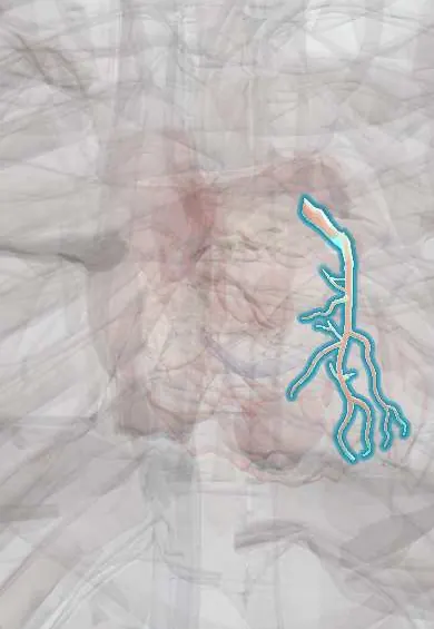
- LAD (anterior descending artery)
- Circumflex artery
- Right coronary artery
- Left main coronary artery
- Left ventricle
- Posterior descending artery (PDA)
In biochemistry
In the clinical exam
- Chest pain: ischemic, pericardial, pleural and other patterns
- Analysing a symptom: onset, site, character, radiation and more
In physiology
- The ECG in ischemia and infarction
- Coronary circulation and myocardial ischemia
- Muscle blood flow and the cardiovascular response to exercise
- Exercise and training physiology
Why it happens
What it is
Predictable chest pain or discomfort brought on by exertion or emotion and relieved by rest or nitrates, caused by temporary myocardial ischaemia from fixed coronary narrowing.
Who gets it
Coronary artery disease is a leading cause of death in Colombia and worldwide; stable angina is a common first presentation in middle-aged and older adults.
Causes
- Atherosclerotic plaque narrowing one or more coronary arteries
- Less often: aortic stenosis, hypertrophic cardiomyopathy, coronary spasm or microvascular disease
Risk factors
- Smoking
- Hypertension
- Dyslipidaemia
- Diabetes
- Obesity and physical inactivity
- Family history of early coronary disease
- Older age, male sex
Pathophysiology
A fixed plaque limits how much coronary flow can rise. At rest supply is enough, but when exertion raises heart rate, contractility and wall stress, oxygen demand exceeds supply and ischaemia causes pain. Resting or nitrates lower demand and the pain settles within minutes.
Classification
- Typical angina: substernal discomfort, brought on by exertion or stress, relieved by rest or nitrates
- Atypical angina: two of the three features; non-anginal pain: one or none
- Canadian Cardiovascular Society (CCS) grades I–IV by the activity that causes pain
Symptoms and signs
- Pressure, tightness or heaviness behind the sternum
- Radiation to the left arm, neck, jaw or back
- Brought on by exertion, cold, meals or emotion
- Lasts a few minutes and eases with rest or nitrates
- Breathlessness or fatigue as an angina equivalent (women, older adults, diabetes)
Diagnosis
- Clinical
- History of the pain and pre-test probability of coronary disease; risk factors and examination for valve disease and vascular disease.
- Tests
- Resting ECG (often normal), lipids, glucose or HbA1c, kidney function and blood count. Exercise ECG testing when the ECG allows; troponin when an acute syndrome is suspected.
- Imaging
- Stress imaging (stress echocardiography, nuclear perfusion or stress MRI) or coronary CT angiography to show ischaemia or plaque. Invasive coronary angiography defines anatomy before revascularisation; echo checks ventricular and valve function.
Differential diagnosis
- Acute coronary syndrome
- Gastro-oesophageal reflux or oesophageal spasm
- Musculoskeletal chest wall pain
- Pericarditis
- Pulmonary embolism
- Aortic stenosis
Goal of treatment
Relieve angina and improve quality of life, and prevent myocardial infarction and death by controlling atherosclerosis.
Treatment
- Medical
- Symptom relief: short-acting nitrates for attacks; beta-blockers or calcium channel blockers first, with long-acting nitrates or other anti-anginal drugs added if needed.
- Medical
- Event prevention: antiplatelet therapy, high-intensity statins and, in many patients, ACE inhibitors; control of blood pressure and diabetes.
- Procedure
- Percutaneous coronary intervention with a stent for persistent symptoms despite medication or high-risk anatomy.
- Surgical
- Coronary artery bypass grafting for left main or multivessel disease, especially with diabetes or reduced ventricular function.
- Lifestyle
- Stopping smoking, healthy diet, weight control and regular physical activity.
- Rehabilitation
- Cardiac rehabilitation with supervised exercise.
Drugs for this condition
Complications
- Acute coronary syndrome and myocardial infarction
- Heart failure from ischaemic cardiomyopathy
- Arrhythmias and sudden death
Prognosis
With risk-factor control and treatment, many patients stay stable for years. Prognosis is worse with left main or multivessel disease, reduced ejection fraction or large ischaemic areas on stress testing.
Prevention
- Not smoking
- Control of blood pressure, lipids and diabetes
- Physical activity and a healthy weight
Test yourself
Think of the answer, then tap the question to see it.
What three features define typical angina?
Substernal chest discomfort, brought on by exertion or emotional stress, and relieved by rest or nitrates.
Why does stable angina appear with exertion and not at rest?
A fixed coronary stenosis lets enough flow through at rest, but cannot meet the higher oxygen demand of exercise.
Which test defines coronary anatomy before revascularisation?
Invasive coronary angiography.
When is coronary artery bypass grafting preferred over stenting in stable coronary disease?
Typically in significant left main or multivessel disease, especially with diabetes or reduced left ventricular function.
What distinguishes unstable angina from stable angina?
Unstable angina occurs at rest, is new or is getting worse; it is an acute coronary syndrome needing urgent care.
Related conditions
- Myocardial Infarction (Heart Attack)
- Aortic Valve Stenosis
- Hypertensive heart disease
- Arterial hypertension
- Type 2 diabetes mellitus
- Atherosclerosis and dyslipidaemia
- Pulmonary embolism
- Gastro-oesophageal reflux disease (GERD)
Educational information, not medical advice. For any symptom, see a health professional.
Sources: Stable Angina (StatPearls (NCBI Bookshelf), 2026).
Spotted a mistake?We are always improving for the community. Tell us what is wrong and our team will review it.Report a mistake
Representative model, for educational purposes only.