
Hypophosphatasia
An inherited bone and tooth disease in which an enzyme that hardens bone is missing. A low alkaline phosphatase on a routine blood test is the clue.
Quick facts
- Key signs
- Early loss of primary teeth · Bowed legs
- Diagnosis
- ALPL gene testing
Red flags ↓
No doses: the clinician sets them by the current guideline.
What it is
An inherited disorder of bone and tooth mineralisation caused by pathogenic variants in ALPL, which encodes tissue-nonspecific alkaline phosphatase (TNSALP). It ranges from a lethal perinatal form to adults with stress fractures and early tooth loss: rickets or osteomalacia with a low, not high, alkaline phosphatase.
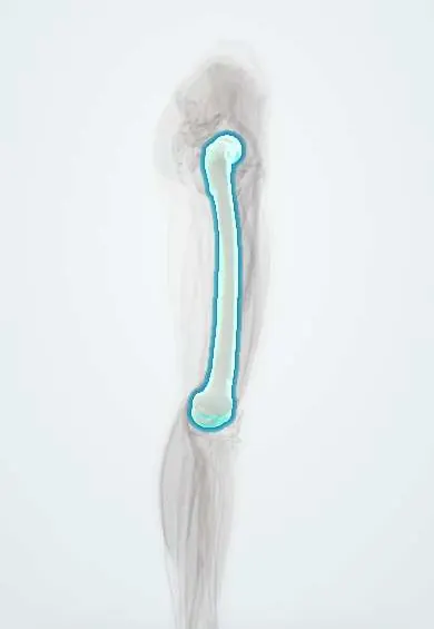
Who gets it
Severe forms are rare (about one in 100 000 to 300 000 births in North America and Europe), with founder populations at much higher risk, such as Mennonites in Manitoba, Canada. Mild forms are far more common, estimated at more than 40 times the severe forms, and many adults are undiagnosed or misdiagnosed for years.
Causes
- Loss-of-function variants in ALPL (chromosome 1), reducing TNSALP activity in bone, liver and kidney
- Severe perinatal and infantile forms: autosomal recessive, with near-absent enzyme activity
- Milder childhood, adult and dental forms: autosomal recessive or dominant (some variants interfere with the normal copy)
- Severity tracks the residual enzyme activity, although the same genotype can vary between people
Risk factors
- Family history of hypophosphatasia, early tooth loss or unexplained fractures
- Consanguinity (severe recessive forms)
- Founder populations (e.g. Manitoba Mennonites)
Pathophysiology
ALPL variant → too little TNSALP on the surface of osteoblasts, chondrocytes and other cells → its substrates accumulate: inorganic pyrophosphate (a potent inhibitor of hydroxyapatite crystal growth), pyridoxal 5′-phosphate (PLP, the circulating form of vitamin B6) and phosphoethanolamine → excess pyrophosphate blocks mineralisation: rickets in children, osteomalacia in adults, poorly formed cementum so teeth fall out with intact roots, and calcium pyrophosphate crystal deposition in joints → calcium and phosphate cannot enter bone, so in severe infant forms they rise in blood and urine, with hypercalcaemia and nephrocalcinosis → PLP cannot be dephosphorylated to cross into the brain, so severe infants have vitamin B6-responsive seizures. In adults the soft bone breaks under ordinary loading: stress fractures of the metatarsals and lateral femoral pseudofractures that heal poorly.
Classification
- Perinatal (severe): profound hypomineralisation in utero, chest deformity, lung hypoplasia, seizures; often lethal
- Benign prenatal: bowed limbs on fetal ultrasound that improve after birth
- Infantile (onset before six months): failure to thrive, rickets, hypercalcaemia, craniosynostosis, respiratory failure
- Childhood: early loss of primary teeth with intact roots, rickets, short stature, bowed legs, delayed walking
- Adult: stress fractures, femoral pseudofractures, chronic bone and joint pain, chondrocalcinosis, loss of adult teeth
- Odontohypophosphatasia: dental disease only, the mildest and probably most common form
Symptoms and signs
- Early loss of primary teeth (before age three) with whole roots, usually lower incisors first
- Bone pain and fractures that heal slowly, especially metatarsal stress fractures
- Thigh pain from femoral pseudofractures
- Bowed legs, widened wrists and ankles and short stature in children
- Muscle weakness and a waddling gait
- Joint pain and inflammation from crystal deposition
- In severe infants: poor feeding, failure to thrive, breathing difficulty and seizures
Diagnosis
- Clinical
- Suspect it in early loss of primary teeth with intact roots, recurrent metatarsal stress fractures, lateral femoral pseudofractures, rickets with a low alkaline phosphatase, or a low ALP found on a routine panel. Ask about family dental and fracture history.
- Tests
- Persistently low serum alkaline phosphatase for age and sex (check that the laboratory flags low values; child ranges are higher). Raised plasma pyridoxal 5′-phosphate (stop vitamin B6 supplements before testing) and raised urinary phosphoethanolamine. Calcium and phosphate are normal or high and PTH normal or low, unlike other rickets. ALPL gene testing confirms; a negative test does not fully exclude it. Exclude other causes of low ALP: drugs, hypothyroidism, zinc or magnesium deficiency, malnutrition, Wilson disease and others.
- Imaging
- Radiographs: rachitic changes and 'tongues' of radiolucency in children; metatarsal stress fractures and lateral subtrochanteric femoral pseudofractures in adults; chondrocalcinosis. Fetal ultrasound for perinatal forms. Bone density supports but does not make the diagnosis.
Differential diagnosis
- Nutritional or X-linked hypophosphataemic rickets and osteomalacia (alkaline phosphatase high)
- Osteoporosis (normal alkaline phosphatase; treated with bisphosphonates, which harm hypophosphatasia)
- Osteogenesis imperfecta (blue sclerae, many fractures, normal ALP)
- Periodontitis or neutrophil disorders causing early tooth loss
- Other causes of a low ALP: drugs, hypothyroidism, zinc or magnesium deficiency, Wilson disease
- Child abuse (unexplained fractures in an infant)
Soft bones and fractures: the alkaline phosphatase sorts them
| Condition | Alkaline phosphatase | Calcium, phosphate, PTH | Key clue | Bisphosphonates |
|---|---|---|---|---|
| Hypophosphatasia | Low | Calcium and phosphate normal or high; PTH normal or low | Teeth lost with roots, high PLP, lateral femoral pseudofractures | Avoid: can worsen fractures |
| Vitamin D-deficiency rickets or osteomalacia | High | Calcium low-normal, phosphate low, PTH high | Low vitamin D, poor sun or diet, malabsorption | Not the treatment; replace vitamin D and calcium |
| Osteoporosis | Normal | Normal | Fragility fractures, low bone density, older age | First-line treatment |
| Osteogenesis imperfecta | Normal | Normal | Blue sclerae, many fractures from childhood, hearing loss | Used in selected children |
Goal of treatment
Restore mineralisation in severe forms with enzyme replacement, and in milder forms control pain, heal fractures, protect the teeth and avoid treatments that block mineralisation.
Treatment
- Medical
- Enzyme replacement with a bone-targeted recombinant TNSALP (asfotase alfa), given by subcutaneous injection; it improves survival, skeletal mineralisation, breathing and motor development in perinatal and infantile forms and is also used in selected children and adults with significant disease. Injection-site reactions are the most common adverse effect.
- Medical
- Avoid bisphosphonates (pyrophosphate analogues that further block mineralisation and can increase fractures) and avoid routine calcium or vitamin D supplements in patients with normal calcium, which can cause hypercalcaemia and hypercalciuria. Anti-inflammatory drugs for bone pain and crystal arthritis; vitamin B6 for seizures in severe infants.
- Acute
- Respiratory support and treatment of hypercalcaemia in severe infants.
- Surgical
- Fixation of non-healing femoral pseudofractures and fractures; neurosurgery for craniosynostosis with raised intracranial pressure.
- Follow-up
- Regular dental care from early childhood, physiotherapy and pain management, and follow-up of growth, mobility, kidney calcification and fracture healing; genetic counselling for the family.
Complications
- Respiratory failure from chest deformity and lung hypoplasia (perinatal and infantile)
- Vitamin B6-responsive seizures in severe infants
- Craniosynostosis and raised intracranial pressure
- Hypercalcaemia, hypercalciuria and nephrocalcinosis
- Recurrent, poorly healing fractures and chronic pain
- Premature loss of primary and permanent teeth
- Chondrocalcinosis and calcium pyrophosphate arthritis
Common traps
- A low alkaline phosphatase is a reassuring result: a persistently low ALP is the key clue to hypophosphatasia and is often ignored because laboratories flag only high values
- Every adult with fractures and low bone density has osteoporosis: in hypophosphatasia bisphosphonates, which are pyrophosphate analogues, add to the block on mineralisation and can cause more fractures
- All rickets has a high alkaline phosphatase: hypophosphatasia is the exception, with low ALP and normal or high calcium and phosphate
- Early loss of a baby tooth is always normal: a primary tooth lost before age three with its whole root, without trauma, points to defective cementum
- Giving calcium and vitamin D will help the soft bones: the problem is not a lack of mineral but a block on depositing it, so supplements can cause hypercalcaemia
Prognosis
The perinatal and infantile forms were often fatal from respiratory failure; enzyme replacement has greatly improved survival and development. Childhood and adult forms are usually compatible with a normal lifespan, but recurrent fractures, pain, muscle weakness and tooth loss can be disabling.
Prevention
- Genetic counselling and, in known families, prenatal diagnosis
- Checking alkaline phosphatase before starting a bisphosphonate
- Early dental care in affected children
Test yourself
Think of the answer, then tap the question to see it.
Which routine blood test result is the main clue to hypophosphatasia?
A persistently low serum alkaline phosphatase for age and sex, unlike the high ALP of other rickets and osteomalacia.
Why are bisphosphonates harmful in hypophosphatasia?
They are pyrophosphate analogues, so they add to the excess pyrophosphate that already blocks bone mineralisation, and can increase fractures.
Why do severely affected infants have vitamin B6-responsive seizures?
Without TNSALP, PLP cannot be dephosphorylated to cross into the brain, so brain vitamin B6 is lacking despite high blood PLP.
Which dental finding suggests hypophosphatasia in a toddler?
Early loss of primary teeth, typically lower incisors before age three, with the entire root intact because cementum did not form.
Where do femoral pseudofractures typically occur in adult hypophosphatasia?
In the lateral subtrochanteric region of the femur, not the femoral neck as in osteomalacia.
Connections
Educational information, not medical advice. For any symptom, see a health professional.
Sources: Hypophosphatasia (MedlinePlus Genetics, US National Library of Medicine, 2026); Hypophosphatasia: a unique disorder of bone mineralization (International Journal of Molecular Sciences) (Europe PMC, 2021).
Spotted a mistake?We are always improving for the community. Tell us what is wrong and our team will review it.Report a mistake
Representative model, for educational purposes only.