
Fracture management principles: describe, reduce, hold, rehabilitate
Bone heals itself if the pieces are lined up and held still long enough without harming the tissue around them. Some injuries cannot wait even an hour.
What it is
The general approach to any fracture: assess the whole patient and the limb, describe the fracture precisely, reduce it when displacement matters, hold the reduction with a splint, cast, traction or fixation, and rehabilitate the limb, while treating the time-critical injuries (open fracture, arterial injury, compartment syndrome, dislocation) first.
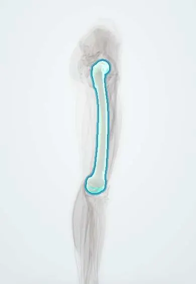
Who gets it
Fractures are among the commonest reasons for emergency visits worldwide. Young adults break bones in high-energy injuries such as road traffic crashes, especially motorcycle crashes in Colombia, while older adults break them in falls from standing because of osteoporosis. Wrist, hip, ankle and shaft fractures of the forearm and leg are the most frequent.
Causes
- High-energy trauma: road traffic crashes, falls from height, crush and gunshot injuries
- Low-energy falls in people with osteoporosis (fragility fractures)
- Repetitive loading (stress fractures)
- Bone weakened by tumour, infection or metabolic disease (pathological fractures)
- Sports injuries and twisting injuries
Risk factors
- Older age, osteoporosis and falls
- Motorcycle riding and other high-speed transport
- Alcohol and drug use
- Contact sports
- Smoking, diabetes and poor nutrition (slower healing)
Pathophysiology
Bone breaks → bleeding forms a fracture haematoma and the inflammatory phase begins, bringing cells that clean up and signal repair → in a fracture with some movement (relative stability, as with a cast or nail), a soft cartilage callus bridges the gap and then mineralises into hard callus over weeks → remodelling reshapes the bone over months to years along the lines of load. In a fracture held rigidly with fragments compressed together (absolute stability, as with a compression plate), bone heals directly across the gap without visible callus. Too much movement or a large gap → the callus cannot bridge and the fracture fails to unite; too little blood supply (scaphoid, femoral neck, talus), infection, smoking or stripping of the soft tissues → the same. Meanwhile the swelling of the injured soft tissues inside closed fascial compartments or a tight cast can raise pressure until it cuts off capillary flow (compartment syndrome).
Classification
- Describe: open or closed; which bone and where (diaphysis, metaphysis, epiphysis, intra-articular); pattern (transverse, oblique, spiral, comminuted, segmental, avulsion, impacted); displacement (translation, angulation, rotation, shortening); neurovascular and skin state
- In children: greenstick, buckle (torus) and physeal fractures, graded by Salter–Harris I–V
- Open fractures by the Gustilo–Anderson grade (I–IIIC); soft tissue in closed fractures by the Tscherne grade; the AO/OTA code for the bone and pattern
- Ways to hold a fracture: splint or backslab (not circumferential, allows swelling), cast (circumferential, once swelling has settled), functional brace, traction (skin or skeletal; a traction splint before hospital for mid-shaft femur fractures), external fixator, internal fixation (plates and screws, intramedullary nails, wires) and joint replacement
Symptoms and signs
- Pain, swelling and bruising after injury
- Deformity, shortening or abnormal rotation of the limb
- Loss of function and inability to bear weight
- Crepitus or abnormal movement at the fracture site
- A wound over or near the fracture (possible open fracture)
- Numbness, weakness, coldness or pallor beyond the injury (nerve or vascular injury)
Diagnosis
- Clinical
- In major trauma, follow ABCDE first; fractures come in the secondary survey unless they are bleeding catastrophically. Then for the limb: look (deformity, wounds, swelling, skin colour), feel (tenderness, pulses, capillary refill, sensation, compartments), move (active movement of fingers or toes, pain on passive stretch), and examine the joint above and below. Record neurovascular status before and after every reduction or splint.
- Tests
- Usually none are needed for the fracture itself. In major or pelvic trauma: haemoglobin, blood group and crossmatch, coagulation and lactate. Before surgery: tests guided by age and illnesses. Compartment pressure measurement when the clinical picture is unclear, for example in an unconscious patient.
- Imaging
- X-rays in two planes at right angles, including the joints above and below the fracture; repeat after every reduction. CT for intra-articular fractures and pelvic, spinal and complex fractures, to plan surgery. MRI for occult fractures (scaphoid, femoral neck) with normal X-rays. CT angiography when an arterial injury is suspected but the limb is not obviously ischaemic.
Differential diagnosis
- Dislocation or fracture-dislocation
- Ligament sprain or tendon rupture
- Soft tissue contusion
- Pathological fracture through a lesion
- Non-accidental injury in a child or older adult
- Septic arthritis or osteomyelitis presenting with pain and swelling
Fracture emergencies: what cannot wait
| Emergency | Clue | Why it is urgent | First move |
|---|---|---|---|
| Open fracture | Wound at or near the fracture, even a small puncture | Bacteria reach the bone; infection risk rises with delay | Early intravenous antibiotics, tetanus cover, sterile dressing, splint; debridement in theatre |
| Compartment syndrome | Pain out of proportion and on passive stretch, tense compartment; pulses usually present | Muscle and nerve die within hours of high pressure | Split the cast and padding to the skin; urgent fasciotomy |
| Arterial injury | Cold, pale, pulseless limb; expanding haematoma | Muscle tolerates warm ischaemia for only a few hours | Realign and splint, recheck pulses; urgent vascular repair or a temporary shunt |
| Dislocation or tented skin | Joint deformity; white, stretched skin over a fragment | The joint's blood supply and the skin are failing | Reduce promptly under analgesia or sedation, then splint and X-ray |
| Pelvic fracture with shock | Hypotension, pelvic pain, leg length difference | Pelvic veins and arteries bleed into a large space | Pelvic binder at the level of the greater trochanters, blood, angiography or packing |
Goal of treatment
Save life and limb first, then obtain a healed bone in an acceptable position with a functional limb: restore length, alignment and rotation, restore joint surfaces exactly, hold them stably enough for healing, and keep muscles and joints moving.
Treatment
- Acute
- First aid and emergency care: analgesia (including regional nerve blocks), realignment of grossly deformed limbs with gentle traction, splinting that includes the joints above and below, elevation, removal of rings and tight items, and neurovascular checks before and after.
- Procedure
- Closed reduction under analgesia, regional block or sedation: traction to restore length, then reverse the deforming force; check X-rays. Rotation must always be corrected; some angulation is acceptable in shaft fractures, more in children; joint surfaces need near-anatomical reduction.
- Conservative
- Non-operative holding: a splint first while swelling settles, then a cast or functional brace for stable, acceptably aligned fractures, with repeat X-rays to detect slipping.
- Procedure
- Traction: a traction splint before hospital for mid-shaft femur fractures (not with pelvic, hip, knee or ankle injury on that side), and skin or skeletal traction in hospital as a temporary measure to control length, pain and bleeding.
- Surgical
- Operative fixation when closed methods fail or are unsuitable: unstable or intra-articular fractures, open fractures, fractures with vascular injury, multiple injuries and pathological fractures. Plates and screws for exact joint reduction, intramedullary nails for long-bone shafts, external fixators for contaminated wounds or unstable patients (damage control orthopaedics), and joint replacement for some femoral neck fractures in older adults.
- Medical
- Venous thromboembolism prophylaxis in lower limb and pelvic fractures with reduced mobility; antibiotics for open fractures; treatment of osteoporosis after a fragility fracture.
- Rehabilitation
- Early movement of free joints, physiotherapy and progressive weight-bearing as the fixation allows, to prevent stiffness, muscle wasting, pressure sores and loss of function.
- Follow-up
- Nursing checks of colour, warmth, capillary refill, movement, sensation and pain on passive stretch, especially in the first day after injury, casting or surgery; urgent review of any worsening pain under a cast.
Complications
- Compartment syndrome and Volkmann ischaemic contracture
- Arterial and nerve injury
- Fat embolism syndrome
- Deep vein thrombosis and pulmonary embolism
- Infection and osteomyelitis
- Delayed union, non-union and malunion
- Avascular necrosis (femoral head, scaphoid, talus)
- Joint stiffness and post-traumatic arthritis
- Complex regional pain syndrome
- Growth disturbance after physeal injury in children
Common traps
- A palpable pulse rules out compartment syndrome: pressure high enough to kill muscle is usually still below arterial pressure; the pulse disappears late, so rely on pain out of proportion and pain on passive stretch
- A circumferential cast is the best first splint: swelling peaks in the first days; use a splint or backslab first and split any cast at once if pain increases
- If the angulation looks fine on X-ray, the alignment is acceptable: rotation is easily missed on X-ray and never remodels; check finger cascade or foot position clinically
- A small wound over a fracture is just a graze: any wound connecting with the fracture makes it open and needs early antibiotics and debridement
- Every fracture needs an operation as soon as possible: the urgent work is skin, vessels, compartments and dislocations; many fractures do well in a splint or cast, and swollen limbs may need staged fixation
Prognosis
Most fractures heal well: upper limb fractures in adults typically unite in about 6–8 weeks and lower limb shafts in about 3–4 months, faster in children. Outcome worsens with open and high-energy injuries, joint involvement, poor blood supply, infection, smoking and diabetes, and when a time-critical injury is missed.
Prevention
- Helmets, protective gear and road safety measures for motorcyclists
- Fall prevention and osteoporosis treatment in older adults
- Workplace safety and fall protection
- Correct cast and splint technique with written warning signs for patients
- Stopping smoking to improve healing
Test yourself
Think of the answer, then tap the question to see it.
Six hours after a tibial shaft fracture is put in a long leg cast, the patient has severe pain made worse by passively moving the toes, but the foot pulses are present. What next?
Suspect compartment syndrome: split the cast and padding down to the skin and elevate to heart level only; if not relieved, urgent fasciotomy. Present pulses do not exclude it.
Why must rotational malalignment always be corrected, even when some angulation is accepted?
Remodelling can straighten some angulation, especially in children and near joints, but rotation does not remodel; it leaves a permanently twisted limb or overlapping fingers.
What is the difference between absolute and relative stability, and how does each heal?
Absolute stability (fragments compressed rigidly, as with a compression plate) heals directly without callus. Relative stability (controlled movement, as with a cast or nail) heals through callus.
Why is a traction splint used for a mid-shaft femur fracture before hospital, and when should it not be used?
It restores length, reduces pain and limits bleeding into the thigh. It is avoided with a pelvic, hip, knee or ankle injury on that side, where pulling would worsen the damage.
Why it happens
In rehabilitation
Connections
Educational information, not medical advice. For any symptom, see a health professional.
Sources: Fracture Healing Overview (StatPearls (NCBI Bookshelf), 2026); Splinting (StatPearls (NCBI Bookshelf), 2023); Acute Compartment Syndrome (StatPearls (NCBI Bookshelf), 2023); Open Fracture Management (StatPearls (NCBI Bookshelf), 2023); EMS Traction Splint (StatPearls (NCBI Bookshelf), 2023).
Spotted a mistake?We are always improving for the community. Tell us what is wrong and our team will review it.Report a mistake
Representative model, for educational purposes only.