Conditions · Gastroenterology and hepatology
Ischaemic colitis !
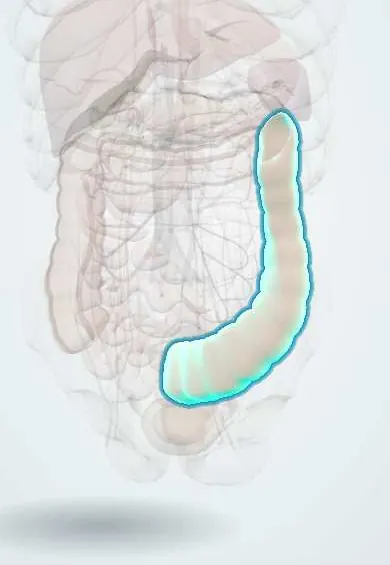
Injury to the colon from a brief drop in its blood supply. It usually causes sudden cramping left-sided belly pain, then bloody diarrhoea, in older adults.
Updated 2026-11-01
Structures involved
Tap one to see what it is and open it in 3D.
- Descending colon
- Sigmoid colon
- Inferior mesenteric artery
- Transverse colon
- Superior mesenteric artery
- Abdominal aorta
- Rectum
- Ascending colon
In the clinical exam
Why it happens
- Infarction: red vs white, and what decides the damage
- Hypoxia, ischemia and reperfusion injury
- Watershed areas and the organs most sensitive to ischemia
- Splanchnic and portal circulation
- Large intestine, appendix and anal canal
What it is
The most common form of intestinal ischaemia: a usually transient reduction of blood flow to the colon that injures the mucosa. Most cases are non-occlusive and resolve with supportive care, but a minority progress to gangrene.
Who gets it
Mainly affects adults over 60, more often women. It is an important cause of lower gastrointestinal bleeding in older people and is likely under-diagnosed because mild cases resolve quickly.
Causes
- Non-occlusive low-flow states: dehydration, heart failure, shock, sepsis, arrhythmia
- Small-vessel atherosclerosis in older adults
- Ligation or loss of the inferior mesenteric artery after aortic aneurysm repair
- Drugs: vasoconstrictors, some laxatives, cocaine, oestrogens and others
- Thrombophilia, vasculitis and, in younger people, long-distance running
Risk factors
- Age over 60
- Cardiovascular disease, atrial fibrillation and heart failure
- Recent aortic surgery or cardiac bypass
- Hypotension, dehydration or major blood loss
- Chronic kidney disease on dialysis
- Constipation and irritable bowel syndrome
Pathophysiology
The colon has relatively poor collateral flow at the 'watershed' areas between arterial territories: the splenic flexure (Griffiths point, between superior and inferior mesenteric arteries) and the rectosigmoid junction (Sudeck point). When perfusion drops, the mucosa, which is most sensitive, becomes ischaemic first, causing oedema, haemorrhage and ulceration. Restoring flow usually allows healing; transmural ischaemia leads to necrosis and perforation.
Classification
- Non-gangrenous (most cases): transient, reversible colitis or chronic ischaemic colitis with stricture
- Gangrenous: transmural necrosis, a surgical emergency
- Isolated right colon ischaemia: less common, more often severe and may signal superior mesenteric artery disease
Symptoms and signs
- Sudden, mild to moderate cramping pain, usually in the left lower abdomen
- Urge to defecate followed within a day by bright red or maroon blood mixed with stool
- Mild tenderness over the affected segment
- Pain that is milder and less out of proportion than in acute mesenteric ischaemia
Diagnosis
- Clinical
- Suspected in an older adult with sudden left-sided cramping pain followed by bloody diarrhoea, especially after hypotension, aortic surgery or dehydration. Stool studies help exclude infection.
- Tests
- Blood count, electrolytes, kidney function, lactate and coagulation; stool cultures and Clostridioides difficile testing. Colonoscopy with biopsy, done carefully with minimal air insufflation, confirms the diagnosis and shows a segmental pattern with a pale or dusky mucosa, oedema and ulcers.
- Imaging
- Contrast CT shows segmental bowel wall thickening and excludes perforation; 'thumbprinting' may be seen on plain films. Mesenteric angiography is usually not needed unless isolated right colon involvement or acute mesenteric ischaemia is suspected.
Differential diagnosis
- Infectious colitis (including Clostridioides difficile)
- Inflammatory bowel disease
- Diverticular bleeding or diverticulitis
- Acute mesenteric ischaemia
- Colorectal cancer
Goal of treatment
Restore perfusion and support the colon while it heals, treat the cause of low flow, and detect early the minority who develop gangrene or perforation and need surgery.
Treatment
- Conservative
- Bowel rest, intravenous fluids to correct volume and improve perfusion, and stopping vasoconstrictive drugs; correct heart failure or arrhythmia.
- Medical
- Broad-spectrum antibiotics for moderate or severe cases to limit bacterial translocation.
- Follow-up
- Serial abdominal examinations, vital signs, lactate and blood counts to detect worsening; repeat colonoscopy may be used to follow severe cases.
- Surgical
- Resection of the affected colon for gangrene, perforation, peritonitis, massive bleeding, or later a symptomatic stricture.
Complications
- Gangrene and perforation with peritonitis
- Colonic stricture weeks to months later
- Chronic segmental ischaemic colitis
- Recurrence
- Sepsis
Prognosis
Most non-gangrenous cases resolve within days to two weeks with supportive care. Gangrenous colitis and isolated right-sided disease carry a much higher mortality.
Prevention
- Maintaining hydration and blood pressure in older and post-operative patients
- Care with drugs that reduce bowel blood flow
- Managing cardiovascular risk factors
Test yourself
Think of the answer, then tap the question to see it.
Which two colonic watershed areas are most vulnerable to ischaemia?
The splenic flexure (Griffiths point) and the rectosigmoid junction (Sudeck point).
What is the classic presentation of ischaemic colitis?
An older adult with sudden crampy left lower abdominal pain followed within 24 hours by bloody diarrhoea.
Which test confirms ischaemic colitis?
Colonoscopy with biopsy (CT supports the diagnosis and excludes complications).
Which surgery classically precedes ischaemic colitis of the left colon?
Abdominal aortic aneurysm repair with loss of inferior mesenteric artery flow.
What is the first-line management for non-gangrenous ischaemic colitis?
Supportive care: bowel rest, intravenous fluids, treating the low-flow cause, and antibiotics in moderate to severe cases.
Related conditions
- Acute mesenteric ischaemia
- Chronic mesenteric ischemia
- Lower gastrointestinal bleeding
- Diverticular disease and diverticulitis
- Clostridioides difficile infection
- Ulcerative colitis
- Crohn disease
- Atrial fibrillation
- Heart failure
Educational information, not medical advice. For any symptom, see a health professional.
Sources: Ischemic Colitis - Gastroenterology - MSD Manual Professional Edition (MSD Manual Professional Edition, 2025); Mesenteric artery ischemia: MedlinePlus Medical Encyclopedia (MedlinePlus (U.S. National Library of Medicine), 2025).
Spotted a mistake?We are always improving for the community. Tell us what is wrong and our team will review it.Report a mistake
Representative model, for educational purposes only.