Conditions · Gastroenterology and hepatology
Lower gastrointestinal bleeding !
Bleeding from the colon, rectum or anus, usually seen as red or maroon blood in the stool. Most episodes stop on their own, but some are life-threatening.
Updated 2026-11-01
Structures involved
Tap one to see what it is and open it in 3D.
- Sigmoid colon
- Descending colon
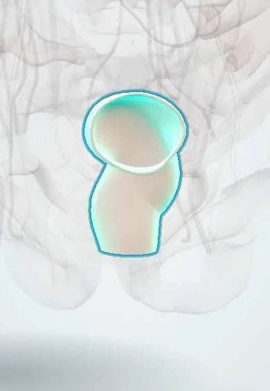
- Rectum
- Ascending colon
- Cecum
- Transverse colon
- Inferior mesenteric artery
- Superior mesenteric artery
In embryology
In the clinical exam
Why it happens
- Hemorrhage: petechiae, purpura, ecchymosis, hematoma, and what blood loss does
- Splanchnic and portal circulation
- Large intestine, appendix and anal canal
- Blood vessels: artery, vein and capillary layers
What it is
Bleeding that originates in the colon, rectum or anus. It usually shows as hematochezia (bright red or maroon blood per rectum) and ranges from minor outlet bleeding to massive haemorrhage with shock.
Who gets it
Less frequent than upper GI bleeding and mainly a disease of older adults, whose risk rises with diverticulosis, vascular disease and the use of antiplatelet drugs and anticoagulants. In Colombia and Latin America, as populations age and colonoscopy access grows, diverticular bleeding and colorectal neoplasia are increasingly recognised causes.
Causes
- Diverticular bleeding: the most common cause in adults; sudden, painless and often large
- Angiodysplasia (vascular ectasia): fragile dilated vessels, typically in the caecum and right colon of older people
- Haemorrhoids and anal fissure: small amounts of bright red blood on the paper or coating the stool
- Colitis: ischaemic, infectious or inflammatory bowel disease, usually with diarrhoea and pain
- Colorectal cancer and polyps, including bleeding after polypectomy
- Radiation proctitis
Risk factors
- Age over 60
- Diverticulosis
- Use of NSAIDs, antiplatelet agents or anticoagulants
- Chronic kidney disease and aortic stenosis (associated with angiodysplasia)
- Previous pelvic radiation or recent polypectomy
Pathophysiology
In diverticular disease the vasa recta stretch over the dome of a diverticulum and can rupture into the lumen, giving brisk arterial bleeding without inflammation. Angiodysplasias are thin-walled ectatic vessels in the submucosa that ooze, often intermittently. Inflammatory and ischaemic colitis bleed from injured mucosa, while tumours bleed from friable surface vessels, often slowly.
Classification
- By severity: minor or self-limited versus severe (haemodynamic instability or need for transfusion)
- By pace: acute overt bleeding versus chronic occult bleeding with iron-deficiency anaemia
- By source: diverticular, vascular, inflammatory, neoplastic or anorectal
Symptoms and signs
- Bright red or maroon blood per rectum (hematochezia)
- Painless bleeding in diverticular disease and angiodysplasia
- Abdominal pain and diarrhoea when the cause is colitis
- Dizziness, fainting, pallor and palpitations when blood loss is large
- Change in bowel habit, weight loss or fatigue when a tumour bleeds slowly
Diagnosis
- Clinical
- Assess haemodynamic status first (pulse, blood pressure, orthostatic changes, urine output). History of drugs, prior bleeding and bowel habit; abdominal and digital rectal examination, and anoscopy for outlet causes.
- Tests
- Complete blood count, coagulation tests, type and cross-match, creatinine and urea; a high urea-to-creatinine ratio suggests an upper GI source. Colonoscopy after bowel preparation, usually within 24 hours of admission, is the main diagnostic and therapeutic test; upper endoscopy first when an upper source is suspected.
- Imaging
- CT angiography locates active bleeding in unstable patients or when colonoscopy is not possible; catheter angiography allows embolisation. Tagged red-cell scintigraphy detects slower intermittent bleeding.
Differential diagnosis
- Upper GI bleeding with rapid transit (peptic ulcer, varices)
- Small-bowel bleeding (angiodysplasia, Meckel diverticulum in young people)
- Red-coloured stool from food such as beetroot or food dyes
- Menstrual or urinary blood mistaken for rectal bleeding
Goal of treatment
Restore and maintain circulating volume, find and stop the bleeding source, and prevent rebleeding, while balancing bleeding against thrombotic risk when antithrombotic drugs are involved.
Treatment
- Acute
- Resuscitation with two large-bore intravenous lines, crystalloids and a restrictive red-cell transfusion strategy; correction of coagulopathy and review of anticoagulants and antiplatelet drugs.
- Procedure
- Colonoscopic haemostasis: clips, thermal coagulation, epinephrine injection or band ligation for diverticular bleeding; argon plasma coagulation for angiodysplasia.
- Procedure
- Selective angiographic embolisation when bleeding is active on CT angiography and endoscopy fails or is not feasible.
- Surgical
- Segmental colectomy for persistent or recurrent bleeding from a localised source; subtotal colectomy is reserved for massive bleeding of unknown site.
- Follow-up
- Nursing care: frequent vital signs, stool count and character, intake and output, serial haemoglobin, and fall precautions because of dizziness.
Complications
- Hypovolaemic shock
- Rebleeding, common in diverticular disease
- Myocardial ischaemia or acute kidney injury from hypoperfusion
- Chronic iron-deficiency anaemia
- Complications of transfusion, angiography (contrast nephropathy, bowel ischaemia) or surgery
Prognosis
Most lower GI bleeding stops spontaneously and mortality is lower than in upper GI bleeding. Older age, comorbidity, anticoagulation and bleeding that starts in hospital predict worse outcomes; diverticular bleeding often recurs.
Prevention
- Avoiding unnecessary NSAIDs in people with prior diverticular bleeding
- Regular review of the need for antithrombotic drugs
- Colorectal cancer screening
Test yourself
Think of the answer, then tap the question to see it.
What is the most common cause of acute lower GI bleeding in adults?
Diverticular bleeding: sudden, painless and often large-volume hematochezia.
Which cause of lower GI bleeding is linked to aortic stenosis and chronic kidney disease?
Angiodysplasia (vascular ectasia), typically in the caecum and right colon of older adults.
Why must an upper source be considered in hematochezia with haemodynamic instability?
A brisk upper GI bleed can pass quickly as red or maroon stool; a high urea-to-creatinine ratio supports it, and upper endoscopy is done first.
What is the first-line test to find and treat lower GI bleeding in a stable patient?
Colonoscopy after bowel preparation, usually within 24 hours.
Which imaging test is preferred for active bleeding in an unstable patient?
CT angiography, followed by catheter angiography with embolisation if it shows active extravasation.
Related conditions
- Upper gastrointestinal bleeding
- Diverticular disease and diverticulitis
- Colorectal cancer
- Haemorrhoids
- Ulcerative colitis
- Crohn disease
- Acute mesenteric ischaemia
- Iron deficiency anaemia
- Hypovolaemic shock
Educational information, not medical advice. For any symptom, see a health professional.
Sources: Symptoms & Causes of GI Bleeding - NIDDK (National Institute of Diabetes and Digestive and Kidney Diseases (NIDDK), 2024); GI Bleed | Gastrointestinal Bleeding: MedlinePlus (MedlinePlus (U.S. National Library of Medicine), 2024).
Spotted a mistake?We are always improving for the community. Tell us what is wrong and our team will review it.Report a mistake
Representative model, for educational purposes only.