Peripartum cardiomyopathy !
Heart failure from a weak, enlarged heart that starts late in pregnancy or in the months after birth, with no other cause found.
Updated 2026-11-01
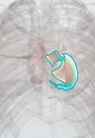
Structures involved
Tap one to see what it is and open it in 3D.
- Left ventricle
- Left atrium
- Mitral anterior leaflet
- Mitral posterior leaflet
- Pulmonary veins
- Interventricular septum
Why it happens
- Maternal physiological changes in pregnancy
- Heart failure physiology
- Prolactin and its control by dopamine
- Reactive oxygen species and antioxidants
What it is
Peripartum cardiomyopathy is heart failure due to left ventricular systolic dysfunction (ejection fraction usually below 45 %) that develops towards the end of pregnancy or in the months after delivery, in a woman without another identifiable cause. It behaves as a dilated cardiomyopathy, although the ventricle is not always dilated. It is an important cause of maternal heart failure and maternal death from cardiovascular disease.
Who gets it
Incidence varies widely between regions, being higher in parts of Africa, Haiti and South Asia and in women of African ancestry. It is a recognised cause of maternal morbidity in Latin America, and Colombia includes heart disease among the conditions reviewed in surveillance of extreme maternal morbidity. Most cases are diagnosed in the first month after delivery.
Causes
- Vascular-hormonal injury: a cleaved fragment of prolactin with anti-angiogenic, cardiotoxic effects, and placental anti-angiogenic factors
- Genetic predisposition (variants in genes for dilated cardiomyopathy, such as titin)
- Inflammation and oxidative stress
- Unmasking of a previously silent cardiomyopathy
Risk factors
- Preeclampsia and hypertensive disorders of pregnancy (strongest association)
- Multiple pregnancy
- Older maternal age and multiparity
- African ancestry
- Obesity, smoking, alcohol use and poor nutrition
- Previous peripartum cardiomyopathy and family history of cardiomyopathy
Pathophysiology
Late pregnancy and the postpartum period bring oxidative stress that can lead to cleavage of prolactin into a 16 kDa fragment that damages endothelium and cardiomyocytes. Placental anti-angiogenic factors, which are also raised in preeclampsia, add to vascular injury. In a genetically susceptible heart, this tips the balance into contractile failure. The weakened left ventricle dilates, filling pressures rise and pulmonary congestion follows; low output and blood stasis favour thrombus and arrhythmia.
Symptoms and signs
- Shortness of breath on exertion, then when lying flat (orthopnoea) or at night
- Fatigue and reduced exercise tolerance
- Ankle and leg swelling beyond the usual swelling of pregnancy
- Palpitations
- Cough, chest discomfort and frequent night-time urination
Diagnosis
- Clinical
- Many symptoms overlap with normal pregnancy, so delay is common; any new dyspnoea, orthopnoea or tachycardia late in pregnancy or post partum deserves evaluation. Diagnosis needs heart failure with reduced ejection fraction near the end of pregnancy or in the months after delivery, with other causes excluded.
- Tests
- Natriuretic peptides (raised, and not substantially raised by normal pregnancy), ECG (sinus tachycardia, nonspecific changes, arrhythmias), troponin, blood count, kidney and thyroid function, and evaluation for preeclampsia.
- Imaging
- Echocardiography is the key test: reduced left ventricular ejection fraction, often with dilation, and screening for intracardiac thrombus. Chest radiography shows congestion. Cardiac magnetic resonance helps exclude myocarditis and other cardiomyopathies.
Differential diagnosis
- Pre-existing dilated cardiomyopathy or valve disease unmasked by pregnancy
- Preeclampsia with pulmonary oedema
- Pulmonary embolism and amniotic fluid embolism
- Myocarditis and Chagas cardiomyopathy
- Takotsubo cardiomyopathy
- Myocardial infarction (including coronary artery dissection)
Goal of treatment
Relieve congestion, protect the heart for recovery, deliver safely, prevent thromboembolism and plan future pregnancies.
Treatment
- Acute
- Acute heart failure or shock: oxygen, diuretics and vasodilators, inotropes or mechanical support in severe cases, with care in a centre with cardiology, obstetrics and intensive care; urgent delivery is considered in decompensated pregnant women.
- Medical
- During pregnancy: beta-blockers, diuretics and vasodilators (hydralazine with nitrate) chosen for fetal safety; ACE inhibitors, ARBs, mineralocorticoid antagonists and other agents harmful to the fetus are avoided until after delivery.
- Medical
- After delivery: full guideline therapy for reduced ejection fraction, with drugs compatible with breastfeeding when the mother breastfeeds.
- Medical
- Bromocriptine, a dopamine agonist that suppresses prolactin, may be added in selected cases with anticoagulation; anticoagulation also for intracardiac thrombus or atrial fibrillation.
- Procedure
- Wearable defibrillator in the early phase in selected cases; implantable defibrillator, ventricular assist device or heart transplantation when function does not recover.
- Follow-up
- Nursing: daily weight, fluid balance, oxygen saturation, signs of congestion, fetal monitoring when pregnant, thromboprophylaxis, and education on contraception and the risk of recurrence.
Complications
- Acute pulmonary oedema and cardiogenic shock
- Ventricular arrhythmias and sudden death
- Thromboembolism, including stroke
- Persistent heart failure needing transplantation
- Recurrence or deterioration in a later pregnancy
- Maternal death
Prognosis
About half of women recover normal ventricular function, usually within six to twelve months; recovery is less likely with a very low ejection fraction or a very dilated ventricle. A subsequent pregnancy carries a significant risk of relapse and death, especially if function has not recovered.
Prevention
- Prenatal care with early detection and control of hypertensive disorders of pregnancy
- Counselling and effective contraception for women with a history of the condition
- Echocardiographic assessment before any future pregnancy
Test yourself
Think of the answer, then tap the question to see it.
When does peripartum cardiomyopathy appear?
Towards the end of pregnancy or in the months after delivery, with no other cause of heart failure.
Which pregnancy disorder is most strongly associated with it?
Preeclampsia and other hypertensive disorders of pregnancy.
Which heart failure drug classes are avoided during pregnancy?
ACE inhibitors, ARBs and mineralocorticoid antagonists (fetal toxicity).
Which hormone is implicated in the pathophysiology?
Prolactin, through a cleaved fragment that injures endothelium and myocardium.
Why is counselling about future pregnancy essential?
Because a subsequent pregnancy carries a significant risk of relapse and death, especially if function has not recovered.
Related conditions
- Preeclampsia
- HELLP syndrome
- Multiple pregnancy and twin–twin transfusion syndrome
- Dilated cardiomyopathy
- Heart failure
- Myocarditis
- Pulmonary embolism
- Amniotic fluid embolism
- Ventricular tachycardia
- Ischaemic stroke
- Cardiogenic shock
- Obesity
- Chagas cardiomyopathy
Educational information, not medical advice. For any symptom, see a health professional.
Sources: Peripartum cardiomyopathy: MedlinePlus Medical Encyclopedia (MedlinePlus, US National Library of Medicine, 2026); Cardiomyopathy - Types | NHLBI, NIH (National Heart, Lung, and Blood Institute (NHLBI), NIH, 2026).
Spotted a mistake?We are always improving for the community. Tell us what is wrong and our team will review it.Report a mistake
Representative model, for educational purposes only.