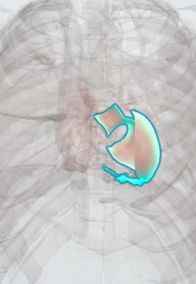
Myocarditis !
Myocarditis is inflammation of the heart muscle, usually after a viral infection. It can cause chest pain, heart failure or dangerous heart rhythms.
Updated 2026-11-01
Structures involved
Tap one to see what it is and open it in 3D.
- Left ventricle
- Right ventricle
- Pericardium
- Bundle branches
- Atrioventricular (AV) node
- Interventricular septum
Why it happens
- When leukocytes injure tissue: proteases, oxidants and NETs
- Patterns of necrosis: coagulative, liquefactive, caseous, fat, fibrinoid, gangrenous
- Heart failure physiology
- How arrhythmias arise
- Cardiac muscle: intercalated discs
What it is
Inflammatory disease of the myocardium, most often triggered by a viral infection, with myocyte injury that is not due to coronary ischaemia. It ranges from silent forms to fulminant heart failure, and can evolve to dilated cardiomyopathy.
Who gets it
Probably underdiagnosed because many cases are mild. It mainly affects young adults and is more common in men; it is an important cause of sudden death in young people and athletes. In Colombia and Latin America, acute Chagas disease (including oral outbreaks from contaminated juices) causes myocarditis, and viral infections such as COVID-19, influenza and enteroviruses are frequent triggers.
Causes
- Viruses: enteroviruses (coxsackie B), adenovirus, parvovirus B19, human herpesvirus 6, influenza, SARS-CoV-2, HIV
- Parasites and bacteria: Trypanosoma cruzi (Chagas disease), diphtheria toxin, Borrelia (Lyme disease)
- Autoimmune and systemic diseases: systemic lupus erythematosus, sarcoidosis, giant cell myocarditis
- Drugs and toxins: immune checkpoint inhibitors, some antipsychotics and anticancer agents, cocaine, alcohol
- Hypersensitivity (eosinophilic) reactions and, rarely, after mRNA vaccination, usually mild
Risk factors
- Recent viral respiratory or gastrointestinal infection
- Young male sex
- Living in or coming from areas with Chagas disease transmission
- Autoimmune disease or immunosuppression
- Treatment with immune checkpoint inhibitors
Pathophysiology
The agent damages myocytes directly, and the immune response (T lymphocytes, cytokines, antibodies against cardiac proteins) amplifies the injury. Inflammation and oedema reduce contractility and disturb conduction, causing heart failure and arrhythmias. Most patients heal, but persistent inflammation or extensive necrosis leaves fibrosis and a dilated, poorly contracting ventricle.
Classification
- By course: acute, fulminant (with cardiogenic shock), chronic active
- By histology: lymphocytic, eosinophilic, giant cell, granulomatous (sarcoidosis)
- By cause: infectious, immune-mediated, toxic or drug-related
Symptoms and signs
- Chest pain that can mimic a heart attack, sometimes pleuritic when the pericardium is involved (myopericarditis)
- Dyspnoea, fatigue and reduced exercise tolerance
- Palpitations, syncope or sudden death from arrhythmias
- Preceding fever, myalgia, cough or diarrhoea days to weeks earlier
- Signs of heart failure: tachycardia, S3 gallop, oedema, raised jugular venous pressure
Diagnosis
- Clinical
- Suspect it in a young patient with chest pain, heart failure or arrhythmia shortly after a viral illness, especially with normal coronary arteries. Ask about travel or residence in Chagas areas, drugs and autoimmune disease.
- Tests
- Raised troponin; ECG with ST and T changes, arrhythmias or conduction block (nonspecific); natriuretic peptides; inflammatory markers; serology or PCR for suspected causes (for example Trypanosoma cruzi). Endomyocardial biopsy is the reference standard and is reserved for severe or unexplained cases, especially to detect giant cell myocarditis.
- Imaging
- Echocardiography shows regional or global ventricular dysfunction and pericardial effusion. Cardiac MRI shows oedema and patchy non-ischaemic late gadolinium enhancement. Coronary angiography or CT is used to exclude an acute coronary syndrome.
Differential diagnosis
- Acute coronary syndrome
- Acute pericarditis
- Takotsubo cardiomyopathy
- Dilated cardiomyopathy of other causes
- Pulmonary embolism
Goal of treatment
Support cardiac function, treat heart failure and arrhythmias, treat the specific cause when there is one, and avoid strenuous exercise while the myocardium heals.
Treatment
- Medical
- Standard heart failure therapy when the ejection fraction is reduced: ACE inhibitors or angiotensin receptor blockers, beta-blockers, mineralocorticoid antagonists and diuretics.
- Acute
- Fulminant myocarditis: inotropes, vasopressors and mechanical circulatory support in a specialised centre, as a bridge to recovery or transplant.
- Medical
- Specific treatment by cause: immunosuppression for giant cell, eosinophilic or sarcoid myocarditis, and antiparasitic therapy for acute Chagas disease.
- Procedure
- Temporary pacing for high-grade block; implantable defibrillator in selected patients with persistent life-threatening ventricular arrhythmias.
- Lifestyle
- Avoid competitive sport and strenuous exercise for several months, until reassessment shows recovery; avoid alcohol and non-steroidal anti-inflammatory drugs in the acute phase.
- Follow-up
- Nursing priorities: continuous cardiac monitoring, blood pressure, fluid balance and daily weight, oxygen saturation and early detection of shock or arrhythmias.
Complications
- Heart failure and cardiogenic shock
- Ventricular arrhythmias and sudden cardiac death
- Atrioventricular block
- Dilated cardiomyopathy
- Need for heart transplantation
Prognosis
Most patients with mild disease and preserved function recover fully. Fulminant myocarditis can recover well if the patient survives the acute phase with support. Giant cell myocarditis and persistent dysfunction have a poor outlook, and some patients progress to dilated cardiomyopathy.
Prevention
- Vaccination against influenza, COVID-19 and diphtheria
- Chagas control: vector control in housing, safe food and juice handling, and screening of blood donors and pregnant women
- Rest from strenuous exercise during and after febrile viral illnesses
Test yourself
Think of the answer, then tap the question to see it.
Which virus group is classically associated with viral myocarditis?
Enteroviruses, especially coxsackie B virus.
What is the reference-standard test for myocarditis, and when is it used?
Endomyocardial biopsy, reserved for severe, unexplained or rapidly worsening cases, for example to detect giant cell myocarditis.
Which non-invasive imaging test shows myocardial oedema and non-ischaemic late enhancement?
Cardiac magnetic resonance imaging.
Which parasitic infection, common in Latin America, can cause acute myocarditis?
Chagas disease (Trypanosoma cruzi), including outbreaks from contaminated food and drinks.
Which long-term complication can follow myocarditis?
Dilated cardiomyopathy with heart failure.
Related conditions
- Dilated cardiomyopathy
- Heart failure
- Acute pericarditis
- Chagas disease
- Chagas cardiomyopathy
- COVID-19
- Influenza (flu)
- Ventricular tachycardia
- Cardiac arrest (VF, pulseless VT, asystole, PEA)
- Myocardial Infarction (Heart Attack)
- Cardiogenic shock
- Sarcoidosis
- Systemic lupus erythematosus
- Diphtheria
Educational information, not medical advice. For any symptom, see a health professional.
Sources: Myocarditis: MedlinePlus Medical Encyclopedia (MedlinePlus, US National Library of Medicine, 2026).
Spotted a mistake?We are always improving for the community. Tell us what is wrong and our team will review it.Report a mistake
Representative model, for educational purposes only.