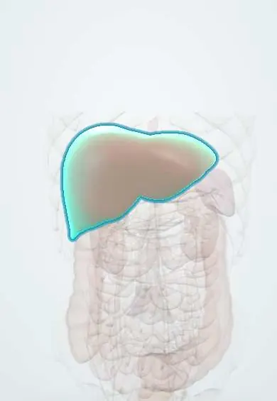
Amoebic liver abscess !
An amoebic liver abscess is a pocket of destroyed liver tissue caused by the parasite Entamoeba histolytica, which travels from the colon to the liver.
Updated 2026-10-29
Structures involved
Tap one to see what it is and open it in 3D.
Microorganisms that cause it
Why it happens
- Patterns of necrosis: coagulative, liquefactive, caseous, fat, fibrinoid, gangrenous
- Liver: lobule, acinus, hepatocytes, sinusoids, Kupffer cells
What it is
A collection of liquefied liver tissue caused by the parasite Entamoeba histolytica, which spreads from the colon to the liver through the portal vein. It is the most common extraintestinal form of amoebiasis.
Who gets it
Occurs where sanitation is poor, including much of Latin America, and in travellers and migrants from these areas. It is much more common in adult men than in women or children; alcohol use is a recognised risk factor.
Causes
- Entamoeba histolytica, acquired by swallowing cysts in contaminated water or food
- Spread of trophozoites from the colon wall through the portal vein to the liver
Risk factors
- Living in or travelling to endemic areas
- Poor sanitation and unsafe drinking water
- Male sex, young to middle adult age
- Alcohol use
- Immunosuppression, malnutrition
Pathophysiology
Trophozoites invade the colonic mucosa and reach the liver via the portal circulation, where they kill hepatocytes and cause liquefactive necrosis. The abscess is usually single and in the right lobe, often near the diaphragm, and contains thick, odourless brown fluid described as anchovy paste. Trophozoites lie at the abscess wall, not in the fluid.
Classification
- Uncomplicated vs complicated (rupture into pleura, peritoneum or pericardium)
- Right-lobe vs left-lobe abscess (left-lobe abscesses risk pericardial rupture)
Symptoms and signs
- Fever, often for days to weeks
- Right upper quadrant pain, sometimes referred to the right shoulder
- Tender enlarged liver; tenderness over the lower right intercostal spaces
- Cough or right pleuritic pain
- Weight loss and malaise; a history of diarrhoea or dysentery in a minority
Diagnosis
- Clinical
- Fever and right upper quadrant pain in a person from or returning from an endemic area, with a tender liver.
- Tests
- Leukocytosis (without eosinophilia), raised alkaline phosphatase and inflammatory markers. Serum antibodies against E. histolytica support the diagnosis but may persist from past infection in endemic areas; antigen tests or PCR where available. Stool microscopy is often negative.
- Imaging
- Ultrasound shows a round, hypoechoic, usually single lesion in the right lobe. CT defines size, location and complications. Aspiration is reserved for selected cases.
Differential diagnosis
- Pyogenic liver abscess
- Hydatid cyst (echinococcosis)
- Necrotic liver tumour or infected cyst
- Acute cholecystitis or cholangitis
- Right lower lobe pneumonia or subphrenic abscess
Goal of treatment
Kill the parasite in the liver and the bowel, relieve symptoms and prevent rupture, using drainage only when it is needed.
Treatment
- Medical
- A nitroimidazole (tissue amoebicide) is the main treatment; most abscesses respond without drainage.
- Medical
- A luminal agent afterwards to clear cysts from the bowel and prevent relapse and transmission.
- Procedure
- Image-guided needle aspiration or catheter drainage for large abscesses, left-lobe abscesses, imminent rupture, no response after several days of treatment or doubt about a pyogenic cause.
- Surgical
- Surgery or drainage of the affected cavity for rupture into the peritoneum, pleura or pericardium.
- Follow-up
- Clinical follow-up of fever and pain; the cavity can take months to resolve on ultrasound, so imaging is not used to judge early response.
Drugs for this condition
Complications
- Rupture into the pleura (empyema) or lung (bronchohepatic fistula)
- Rupture into the peritoneum (peritonitis)
- Rupture into the pericardium (tamponade), mainly from left-lobe abscesses
- Secondary bacterial infection
- Inferior vena cava or hepatic vein compression or thrombosis
Prognosis
With prompt treatment mortality is low and most patients recover fully. Delayed diagnosis and rupture, especially into the pericardium, worsen outcomes.
Prevention
- Safe drinking water and sanitation
- Hand hygiene and safe food handling
- Treatment of carriers with a luminal agent
Test yourself
Think of the answer, then tap the question to see it.
How does Entamoeba histolytica reach the liver?
Trophozoites invade the colonic mucosa and travel through the portal venous system to the liver.
What does the fluid from an amoebic liver abscess look like?
Thick, odourless, reddish-brown fluid often described as anchovy paste; trophozoites are rarely found in it.
Which drug class is the main treatment of amoebic liver abscess, and what follows it?
A nitroimidazole tissue amoebicide, followed by a luminal agent to eradicate intestinal cysts.
Why is a left-lobe amoebic liver abscess considered for drainage?
Because it can rupture into the pericardium and cause tamponade.
Which lobe of the liver is usually affected by an amoebic abscess?
The right lobe, which receives most of the blood from the superior mesenteric vein draining the caecum and right colon.
Related conditions
Educational information, not medical advice. For any symptom, see a health professional.
Sources: Amebic Liver Abscess (StatPearls (NCBI Bookshelf), 2025).
Spotted a mistake?We are always improving for the community. Tell us what is wrong and our team will review it.Report a mistake
Representative model, for educational purposes only.