Conditions · Surgery and trauma
Pulmonary contusion !
A bruise of the lung after a blow to the chest. Bleeding and swelling in the lung tissue make breathing harder over the next one to two days.
Updated 2026-11-01
Structures involved
Tap one to see what it is and open it in 3D.
- Pleura
- Ribs (back part)
- Segmental lung vessels
- Lobar bronchi
- Segmental bronchi
- Sternum
- External intercostals
- Intercostal nerves
- Diaphragm
Why it happens
- Ventilation–perfusion matching and West zones
- The causes of hypoxemia
- Surface tension, surfactant and Laplace's law
- The alveolus: type I and II pneumocytes, surfactant, the blood–air barrier
What it is
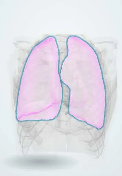
Bruising of the lung parenchyma after blunt chest trauma, with bleeding and oedema in the alveoli and interstitium but no laceration of the lung surface. It is the most common lung injury in blunt chest trauma and often lies under rib fractures or a flail segment.
Who gets it
Found in a large share of patients with significant blunt chest trauma, and commoner in children and young adults, whose flexible chest wall transmits energy to the lung without breaking ribs. In Colombia and Latin America, motorcycle and car crashes, pedestrian injuries and falls from height are the usual mechanisms.
Causes
- Road traffic crashes (motorcycle riders, unrestrained occupants, pedestrians)
- Falls from height
- Crush injuries and direct blows to the chest
- Blast injuries
- High-velocity gunshot wounds (cavitation around the track)
Risk factors
- High-energy mechanism
- Multiple rib fractures or flail chest
- Young age (compliant chest wall)
- Chronic obstructive pulmonary disease or chronic kidney disease (higher need for ventilation)
- Large-volume fluid resuscitation
Pathophysiology
The impact disrupts alveolar capillaries, so blood and fluid fill the alveoli and interstitium. Surfactant loss and local inflammation cause alveolar collapse. The contused area keeps being perfused but is poorly ventilated, producing shunt and ventilation-perfusion mismatch with hypoxaemia. The injury typically worsens over 24–72 hours and then resolves over about a week; large contusions predispose to pneumonia and ARDS.
Classification
- By extent on CT: small versus large (often expressed as the percentage of lung volume involved, with more than about 20% predicting respiratory failure)
- Unilateral versus bilateral
- Isolated versus associated with flail chest, pneumothorax or haemothorax
Symptoms and signs
- Chest pain and chest wall tenderness
- Shortness of breath that may increase gradually
- Tachypnoea and tachycardia
- Cough, sometimes with blood (haemoptysis)
- Reduced breath sounds or crackles over the injured area
Diagnosis
- Clinical
- Suspect it in any patient with blunt chest trauma and breathing difficulty, especially when it worsens over hours. ATLS primary survey, then serial clinical assessment and continuous pulse oximetry.
- Tests
- Arterial blood gas (hypoxaemia, low PaO2/FiO2 ratio, hypercapnia when tiring) and capnometry; baseline trauma blood tests.
- Imaging
- Chest X-ray shows patchy, non-segmental opacities that may not appear for 24–48 hours. CT of the chest is far more sensitive, shows the extent early and detects associated rib fractures, pneumothorax and haemothorax.
Differential diagnosis
- Aspiration pneumonitis or pneumonia
- Haemothorax
- Pulmonary laceration with haematoma
- Fat embolism syndrome
- ARDS from another cause
Goal of treatment
Keep oxygenation and ventilation adequate while the lung heals, control pain so the patient can breathe deeply and cough, and prevent pneumonia and fluid overload.
Treatment
- Acute
- ATLS approach, supplemental oxygen, and treatment of associated pneumothorax or haemothorax with chest drainage.
- Medical
- Effective analgesia (multimodal, including regional techniques such as epidural or nerve blocks in multiple rib fractures) to allow deep breathing and coughing.
- Conservative
- Judicious fluid resuscitation aimed at normal perfusion while avoiding excess fluid; chest physiotherapy, incentive spirometry and early mobilisation.
- Procedure
- Non-invasive ventilation in selected alert patients; intubation and lung-protective mechanical ventilation for respiratory failure. Corticosteroids and prophylactic antibiotics are not used routinely.
- Follow-up
- Nursing priorities: continuous pulse oximetry, respiratory rate and work of breathing, pain scores, fluid balance, positioning, and early signs of pneumonia.
Complications
- Acute respiratory failure
- Pneumonia
- ARDS
- Pneumatocele or lung abscess
- Long-term reduced lung function after large contusions
Prognosis
Most contusions resolve within about 5–7 days. Outcome depends on the size of the contusion and on associated injuries; large or bilateral contusions carry a higher risk of ventilation, pneumonia and ARDS.
Prevention
- Seatbelts, airbags and child restraints
- Motorcycle safety and speed control
- Fall prevention at work and at home
Test yourself
Think of the answer, then tap the question to see it.
Why may the first chest X-ray look normal in a patient with pulmonary contusion?
Opacities often take 24–48 hours to appear on plain film; CT shows the contusion much earlier.
What is the gas-exchange mechanism behind hypoxaemia in pulmonary contusion?
Blood and oedema fill the alveoli, so perfused but poorly ventilated lung causes shunt and V/Q mismatch.
Why is excessive IV fluid avoided in pulmonary contusion?
The injured capillaries leak, so fluid overload worsens alveolar oedema and hypoxaemia; resuscitate to normal perfusion only.
Which two late complications are the main concern after a large pulmonary contusion?
Pneumonia and ARDS.
Related conditions
- Rib fractures
- Flail chest
- Pneumothorax
- Haemothorax
- Acute respiratory distress syndrome (ARDS)
- Major trauma (polytrauma): ATLS primary survey
- Blunt cardiac injury
- Community-acquired pneumonia
- Aspiration pneumonia and pneumonitis
Educational information, not medical advice. For any symptom, see a health professional.
Sources: Pulmonary Contusion - Injuries; Poisoning - Merck Manual Professional Edition (Merck Manual Professional Edition, 2025); Pulmonary contusion and flail chest: an Eastern Association for the Surgery of Trauma practice management guideline (Journal of Trauma and Acute Care Surgery, 2012).
Spotted a mistake?We are always improving for the community. Tell us what is wrong and our team will review it.Report a mistake
Representative model, for educational purposes only.