Conditions · Gastroenterology and hepatology
Pyogenic liver abscess !
A pocket of pus in the liver caused by bacteria, often spreading from the bile ducts or the bowel. It causes fever and right upper abdominal pain.
Updated 2026-11-01
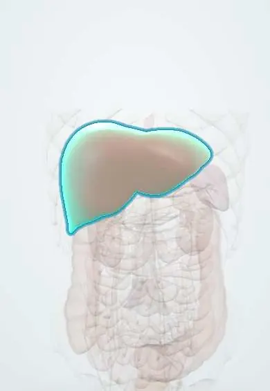
Structures involved
Tap one to see what it is and open it in 3D.
Why it happens
- Outcomes of acute inflammation: resolution, abscess, scar or chronic inflammation
- Patterns of necrosis: coagulative, liquefactive, caseous, fat, fibrinoid, gangrenous
- Splanchnic and portal circulation
- Liver: lobule, acinus, hepatocytes, sinusoids, Kupffer cells
What it is
A collection of pus within the liver caused by bacterial infection. It most often arises from infection of the biliary tree or spreads through the portal vein from an abdominal infection, and it can be single or multiple.
Who gets it
The most common type of liver abscess in high-income countries, mostly in adults over 50. In Colombia and other tropical areas amoebic abscess is also frequent, so both must be considered in every patient with a liver abscess.
Causes
- Biliary: cholangitis from stones, strictures, tumours or after ERCP (most common)
- Portal: appendicitis, diverticulitis, inflammatory bowel disease or bowel perforation
- Arterial: bacteraemia from a distant source such as endocarditis
- Direct spread from the gallbladder or trauma; cryptogenic in many cases
- Organisms: Escherichia coli, Klebsiella pneumoniae, streptococci (Streptococcus anginosus group) and anaerobes, often mixed
Risk factors
- Diabetes mellitus (especially for Klebsiella)
- Biliary disease and previous biliary procedures
- Liver or pancreatic cancer and colorectal cancer
- Older age, immunosuppression and liver transplant
- Chemoembolisation or ablation of liver tumours
Pathophysiology
Bacteria reach the liver through the bile ducts, the portal vein or the hepatic artery. Local infection causes tissue necrosis and a walled-off pus collection, most often in the right lobe because it receives more portal flow. Hypervirulent Klebsiella can spread to the eye, lung and brain.
Classification
- Single versus multiple abscesses
- By route: biliary, portal, arterial, contiguous, cryptogenic
- By size: small (<3–5 cm) versus large
Symptoms and signs
- Fever, chills and night sweats, often the main complaint
- Right upper quadrant pain and tender hepatomegaly
- Malaise, anorexia, nausea and weight loss
- Jaundice, especially with biliary origin
- Right shoulder pain or cough from diaphragmatic irritation
Diagnosis
- Clinical
- Fever with right upper quadrant pain in a patient with biliary or abdominal infection, diabetes or a recent procedure; the classic triad is often incomplete.
- Tests
- Leukocytosis, high CRP, raised alkaline phosphatase and low albumin. Blood cultures (positive in about half) and culture of aspirated pus guide antibiotics. Amoebic serology is done to rule out amoebic abscess, especially in endemic areas such as Colombia.
- Imaging
- Ultrasound is the first test; contrast CT is the most sensitive, shows a rim-enhancing lesion and looks for the source. Colonoscopy is considered in cryptogenic Klebsiella or streptococcal abscess to exclude colorectal cancer.
Differential diagnosis
- Amoebic liver abscess
- Infected hydatid cyst
- Necrotic liver tumour or metastasis
- Acute cholecystitis or cholangitis
- Right lower lobe pneumonia
Goal of treatment
Drain the pus, eradicate the bacteria with prolonged antibiotics and treat the source, while managing sepsis.
Treatment
- Procedure
- Percutaneous drainage guided by ultrasound or CT, by needle aspiration or catheter, for most abscesses larger than about 3–5 cm.
- Medical
- Empirical broad-spectrum antibiotics covering Gram-negative bacteria, streptococci and anaerobes (for example a third-generation cephalosporin with metronidazole, or a beta-lactam/beta-lactamase inhibitor), adjusted to cultures and continued for several weeks.
- Acute
- Sepsis care: blood cultures, early antibiotics, intravenous fluids and close haemodynamic support.
- Surgical
- Surgical drainage or resection for rupture, multiloculated abscesses that fail percutaneous drainage, or when the source needs surgery; ERCP for biliary obstruction.
- Follow-up
- Temperature curve, drain output, inflammatory markers and follow-up imaging until resolution; blood glucose control in diabetes.
Complications
- Sepsis and septic shock
- Rupture into the peritoneum, pleura or pericardium
- Pleural effusion or empyema
- Metastatic infection (endophthalmitis, meningitis) with hypervirulent Klebsiella
- Portal or hepatic vein thrombosis
Prognosis
With drainage and antibiotics, mortality has fallen to under about 10%. Outcome is worse with multiple abscesses, biliary malignancy, shock, older age and delayed diagnosis.
Prevention
- Prompt treatment of biliary and abdominal infections
- Good diabetes control
Test yourself
Think of the answer, then tap the question to see it.
What is the most common source of pyogenic liver abscess?
The biliary tree (cholangitis from stones, strictures or tumours).
Which organism is linked to liver abscess in diabetics and can spread to the eye?
Klebsiella pneumoniae (hypervirulent strains), which can cause endophthalmitis.
Which lobe of the liver is most often involved and why?
The right lobe, because it is larger and receives more portal venous blood.
How does management differ between pyogenic and amoebic liver abscess?
Pyogenic abscess usually needs drainage plus weeks of antibiotics; amoebic abscess usually responds to metronidazole alone.
Why is colonoscopy considered after a cryptogenic Klebsiella or streptococcal liver abscess?
Because it can be the first sign of an underlying colorectal cancer.
Related conditions
- Amoebic liver abscess
- Acute cholangitis
- Choledocholithiasis
- Acute appendicitis
- Diverticular disease and diverticulitis
- Sepsis and septic shock
- Type 2 diabetes mellitus
- Colorectal cancer
Educational information, not medical advice. For any symptom, see a health professional.
Sources: Pyogenic liver abscess: MedlinePlus Medical Encyclopedia (MedlinePlus (U.S. National Library of Medicine), 2024).
Spotted a mistake?We are always improving for the community. Tell us what is wrong and our team will review it.Report a mistake
Representative model, for educational purposes only.