Conditions · Infectious and tropical diseases
Pyomyositis (including psoas abscess) !
Pyomyositis is a bacterial abscess inside a large muscle, usually of the thigh or hip. It is commoner in hot climates and needs drainage and antibiotics.
Updated 2026-11-01
Structures involved
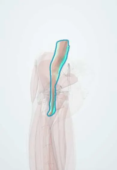
Tap one to see what it is and open it in 3D.
- Psoas major
- Vastus lateralis
- Iliacus
- Rectus femoris
- Vastus medialis
- Gluteus maximus
- Gluteus medius
- Adductor longus
Why it happens
- Outcomes of acute inflammation: resolution, abscess, scar or chronic inflammation
- Skeletal muscle: sarcomere and contraction
What it is
A primary bacterial infection of skeletal muscle that progresses to an intramuscular abscess, usually caused by Staphylococcus aureus. Abscess in the iliopsoas muscle (psoas abscess) may be primary or spread from the spine, bowel or kidney.
Who gets it
Classically a tropical disease (tropical pyomyositis), seen in Africa, Asia, the Pacific and tropical Latin America, including Colombia, and more often in young men and children. It is increasingly reported in temperate countries among people with HIV, diabetes or other immune problems. Psoas abscess in Latin America may still be tuberculous, from spinal tuberculosis (Pott disease).
Causes
- Staphylococcus aureus in most cases, including community-acquired methicillin-resistant strains and strains with Panton-Valentine leukocidin
- Streptococcus pyogenes and, less often, Gram-negative bacilli
- Secondary psoas abscess: spread from vertebral osteomyelitis or spondylodiscitis, Crohn disease, appendicitis, diverticulitis or the kidney (enteric flora)
- Mycobacterium tuberculosis in psoas abscess from Pott disease
Risk factors
- Hot, humid climate and rainy season
- Recent blunt trauma or strenuous use of the muscle
- HIV infection, diabetes, corticosteroid use, cancer or malnutrition
- Staphylococcal skin infection or bacteraemia; injection drug use
- For psoas abscess: spinal infection, inflammatory bowel disease or recent abdominal surgery
Pathophysiology
Healthy muscle resists infection, so a transient bacteraemia usually seeds muscle that has been damaged by trauma or exercise. The infection passes from a diffuse inflammatory (invasive) stage to a collection of pus (suppurative stage) and, if untreated, to sepsis and metastatic abscesses (late stage). The psoas lies close to the lumbar spine, kidney and bowel, so infection there often comes from those neighbours.
Classification
- Stage 1, invasive: firm, woody, painful muscle without pus
- Stage 2, suppurative: abscess with fever, marked pain and swelling (most patients are diagnosed here)
- Stage 3, late: systemic toxicity, septic shock and multiple abscesses
- Psoas abscess: primary (haematogenous) or secondary (contiguous spread)
Symptoms and signs
- Fever with cramping pain in one large muscle, most often the thigh (quadriceps), buttock or calf
- Firm, tender, swollen muscle; overlying skin often only mildly red
- Limp or refusal to bear weight in children
- Psoas abscess: fever, back, flank or groin pain, pain referred to the hip or knee and a hip held in flexion
- Pain on passive hip extension (psoas sign)
Diagnosis
- Clinical
- Muscle pain, swelling and fever in a person from a warm climate or with immune compromise. Early disease is easily mistaken for a strain, haematoma or thrombosis, so suspicion is key.
- Tests
- Leukocytosis and raised CRP or ESR; creatine kinase is often normal, unlike myositis. Blood cultures are positive in a minority. Pus from aspiration or drainage gives the organism and its susceptibility. In psoas abscess, tests look for the source (spinal infection, tuberculosis, bowel disease).
- Imaging
- Ultrasound is a quick first test and guides aspiration. MRI best shows the extent of muscle involvement and abscess. CT with contrast is the most useful test for psoas abscess and shows spinal or abdominal sources.
Differential diagnosis
- Muscle strain, contusion or haematoma
- Deep vein thrombosis
- Cellulitis or necrotising fasciitis
- Septic arthritis of the hip or osteomyelitis
- Soft-tissue sarcoma
- Appendicitis or renal abscess (for psoas abscess)
Goal of treatment
Drain the pus, eradicate the organism and treat the source, preventing sepsis and spread to bone, joints or the spine.
Treatment
- Medical
- Intravenous antistaphylococcal antibiotics chosen by local resistance (anti-MRSA agents where community MRSA is common), broadened to cover enteric bacteria in secondary psoas abscess, then oral step-down; antituberculous therapy for tuberculous psoas abscess.
- Procedure
- Ultrasound- or CT-guided needle aspiration or percutaneous catheter drainage of the abscess.
- Surgical
- Open surgical drainage or debridement when percutaneous drainage fails, collections are multiple, or the source (bowel, spine) needs surgery.
- Follow-up
- Daily review of fever, pain and inflammatory markers; repeat imaging when response is slow; screening for HIV and diabetes.
Complications
- Sepsis, septic shock and toxic shock syndrome
- Metastatic abscesses, endocarditis and pneumonia
- Osteomyelitis and septic arthritis next to the muscle
- Deep vein thrombosis
- Compartment syndrome
- Spinal epidural abscess or recurrence when the source of a psoas abscess is not treated
Prognosis
With early diagnosis, drainage and appropriate antibiotics most people recover fully. Mortality rises with late diagnosis, sepsis, multifocal disease and serious underlying illness.
Test yourself
Think of the answer, then tap the question to see it.
Which organism causes most cases of pyomyositis?
Staphylococcus aureus.
Which muscle group is most often affected in tropical pyomyositis?
The thigh muscles, especially the quadriceps, followed by the gluteal and calf muscles.
What is the best imaging test to diagnose a psoas abscess?
Contrast-enhanced CT, which also shows a spinal or abdominal source.
In Latin America, which chronic infection should be considered with a psoas abscess and back pain?
Spinal tuberculosis (Pott disease), which forms a cold abscess tracking along the psoas.
Is creatine kinase usually high in pyomyositis?
No; it is often normal, which helps distinguish it from inflammatory or viral myositis.
Related conditions
- Osteomyelitis
- Vertebral osteomyelitis
- Spinal tuberculosis (Pott disease)
- Cellulitis of the leg
- Necrotizing soft tissue infection (necrotizing fasciitis, gas gangrene)
- Septic Arthritis of the Knee
- Sepsis and septic shock
- HIV infection and AIDS
- Crohn disease
- Deep vein thrombosis
Educational information, not medical advice. For any symptom, see a health professional.
Sources: Pyomyositis in Children (Current Infectious Disease Reports (PubMed Central), 2016).
Spotted a mistake?We are always improving for the community. Tell us what is wrong and our team will review it.Report a mistake
Representative model, for educational purposes only.