
Subglottic stenosis
Narrowing of the airway just below the vocal cords, inside the cricoid ring. Present at birth or after intubation, it causes stridor and breathlessness.
What it is
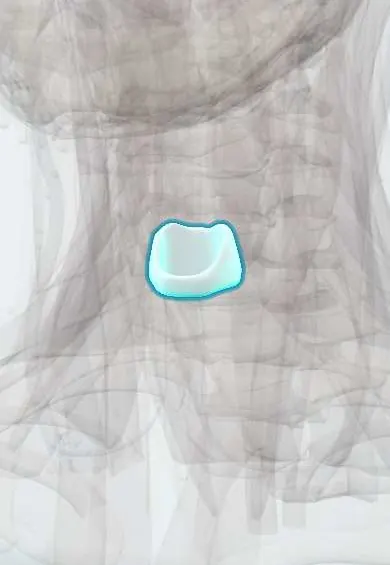
Subglottic stenosis is a narrowing of the airway in the subglottis, the segment below the vocal folds surrounded by the cricoid cartilage, the only complete cartilage ring of the airway. It can be congenital or acquired, most often after prolonged endotracheal intubation.
Who gets it
Most cases are acquired. Prolonged intubation in neonatal intensive care is the leading cause in children, and the condition is now less frequent thanks to better tube care. In adults, intubation and tracheostomy injury predominate; idiopathic subglottic stenosis affects mainly women. In full-term neonates a subglottic diameter under 4 mm (3 mm in premature infants) is considered narrowed.
Causes
- Prolonged or traumatic endotracheal intubation, oversized tubes and high tracheostomy
- Congenital: incomplete canalisation of the larynx or a malformed cricoid
- Autoimmune disease: granulomatosis with polyangiitis, relapsing polychondritis, sarcoidosis
- Idiopathic (mostly women), reflux, trauma, burns, infections and radiation
Risk factors
- Prematurity and long neonatal intubation
- Repeated intubations and tube movement
- Infection or reflux during intubation
- Down syndrome (smaller airway)
- Granulomatosis with polyangiitis
Pathophysiology
Because the cricoid is a rigid complete ring, swelling of the mucosa can only narrow the lumen inward. Tube pressure causes ischaemia, ulceration and perichondritis; healing with granulation tissue and fibrosis leaves a scar that narrows the airway. Airflow resistance rises steeply with small reductions in diameter, so minor swelling from a cold can cause major obstruction.
Classification
- Congenital (membranous or cartilaginous) or acquired
- Cotton-Myer grade by the percentage of lumen obstructed: I up to 50%, II 51 to 70%, III 71 to 99%, IV no detectable lumen
- By length and involvement of the glottis or trachea
Symptoms and signs
- Biphasic stridor or noisy breathing
- Breathlessness on exertion, increasing over time
- Recurrent or prolonged croup-like episodes in a child
- Failed extubation in a neonate
- Chronic cough and difficulty clearing secretions; adults are often misdiagnosed with asthma
Diagnosis
- Clinical
- History of intubation, prematurity or autoimmune disease with biphasic stridor or progressive exertional breathlessness that does not respond to asthma treatment.
- Tests
- Laryngoscopy and bronchoscopy under anaesthesia are the gold standard: they size the airway with endotracheal tubes, give the Cotton-Myer grade and look for other lesions. Flow-volume loops show a fixed upper airway obstruction (flattened inspiratory and expiratory limbs). ANCA and other autoimmune tests when no cause is clear.
- Imaging
- CT of the neck and airway, with 3D reconstruction, measures the length and diameter of the narrowing; airway radiographs may show narrowing in children.
Differential diagnosis
- Croup (acute, self-limited)
- Laryngomalacia and tracheomalacia
- Subglottic haemangioma
- Recurrent respiratory papillomatosis
- Asthma, vocal fold paralysis and inhaled foreign body
Goal of treatment
Secure and widen the airway, allow decannulation or avoid tracheostomy, protect the voice, and treat the cause (reflux, autoimmune disease) to prevent recurrence.
Treatment
- Follow-up
- Mild stenosis (grade I, some grade II) can be observed, as the airway grows in children; treat reflux and any autoimmune disease; refer to an airway team for any symptomatic stenosis.
- Procedure
- Endoscopic treatment: balloon dilation, laser or cold incision of scar and local corticosteroid injection, especially for thin, short and early stenosis; often repeated.
- Surgical
- Open surgery for severe or long stenosis: laryngotracheal reconstruction with cartilage grafts or cricotracheal resection with anastomosis.
- Acute
- Acute respiratory distress: oxygen, nebulised epinephrine and corticosteroids for swelling, and a secure airway (intubation with a smaller tube or tracheostomy) by experienced staff.
Complications
- Acute airway obstruction during respiratory infections
- Tracheostomy dependence and its problems (tube blockage, accidental removal)
- Voice and swallowing changes after surgery
- Restenosis
- Delayed speech development in children with a long-term tracheostomy
Prognosis
Mild stenosis often improves as a child grows. Most children with severe stenosis achieve a safe airway and removal of the tracheostomy after reconstruction, though several procedures may be needed. Idiopathic adult stenosis tends to recur and often needs repeated dilations.
Prevention
- Appropriately sized endotracheal tubes, cuff pressure checks and tube fixation
- Shortest possible intubation and early review of long intubations
Test yourself
Think of the answer, then tap the question to see it.
Why is the subglottis especially prone to narrowing?
It is surrounded by the cricoid cartilage, the only complete cartilage ring of the airway, so swelling can only narrow the lumen.
What is the most common cause of acquired subglottic stenosis in children?
Prolonged endotracheal intubation, often in neonatal intensive care.
Which grading system is used, and what does grade III mean?
The Cotton-Myer system; grade III is 71 to 99% obstruction of the lumen.
What does a flow-volume loop show in subglottic stenosis?
A fixed upper airway obstruction, with flattening of both the inspiratory and expiratory limbs.
Which systemic disease must be considered in an adult with unexplained subglottic stenosis?
Granulomatosis with polyangiitis.
In clinical skills
Connections
Educational information, not medical advice. For any symptom, see a health professional.
Sources: Subglottic Stenosis (StatPearls (NCBI Bookshelf), 2023).
Spotted a mistake?We are always improving for the community. Tell us what is wrong and our team will review it.Report a mistake
Representative model, for educational purposes only.