Conditions · Ophthalmology and ENT
Vocal fold paralysis (recurrent laryngeal nerve injury) !
A vocal fold stops moving because its nerve is damaged, often after thyroid or chest surgery. One side: weak, breathy voice; both: breathing trouble.
Updated 2026-11-01
Structures involved
Tap one to see what it is and open it in 3D.
- Recurrent laryngeal nerves
- Vocal folds
- Thyroid gland
- Vagus nerve
- Superior laryngeal nerve
- Posterior cricoarytenoid
- Lateral cricoarytenoid
- Arytenoid cartilages
- Inferior thyroid artery
- Parathyroid glands
- Trachea
- Esophagus
- Cricothyroid
Why it happens
- Peripheral nerve: endoneurium, perineurium, epineurium; myelin
- Nerve injury and regeneration (Wallerian degeneration)
What it is
Loss of movement of one or both vocal folds due to injury of the vagus nerve or its recurrent laryngeal branch, which supplies all intrinsic laryngeal muscles except the cricothyroid. Unilateral paralysis mainly affects voice and swallowing; bilateral paralysis narrows the airway and can be life-threatening.
Who gets it
Unilateral paralysis is far more common than bilateral. Surgery in the neck and chest (thyroid and parathyroid, anterior cervical spine, carotid, cardiac, oesophageal and lung surgery) is the leading cause in most series, followed by tumours and idiopathic cases. Thyroidectomy, a frequent operation in Colombia and Latin America because of goitre and thyroid cancer, is the classic setting. The left side is affected more often because its nerve runs a longer course into the chest.
Causes
- Surgical injury: thyroidectomy, parathyroidectomy, anterior cervical spine surgery, carotid endarterectomy, cardiac, oesophageal or lung surgery
- Tumours along the nerve course: lung (apex or left hilum), oesophageal, thyroid and mediastinal cancers
- Endotracheal intubation trauma
- Neurological disease: brainstem stroke involving the nucleus ambiguus, motor neurone disease, multiple sclerosis
- Thoracic causes: aortic arch aneurysm or a greatly enlarged left atrium (Ortner syndrome)
- Idiopathic, often presumed viral
Risk factors
- Total thyroidectomy, reoperation, large goitre or thyroid cancer surgery
- Prolonged intubation
- Smoking (lung and oesophageal cancer)
- Previous neck or chest radiotherapy
Pathophysiology
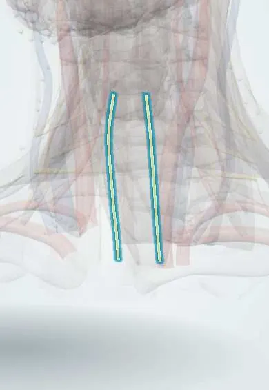
The vagus gives off the recurrent laryngeal nerve, which on the right loops under the subclavian artery and on the left under the aortic arch, then climbs in the tracheo-oesophageal groove close to the inferior thyroid artery and the back of the thyroid. When it is cut, stretched, compressed or burned, the intrinsic muscles on that side stop working: the posterior cricoarytenoid, the only abductor, can no longer open the fold, and the adductors cannot close it. A unilateral paralysed fold rests near the midline but cannot meet its partner, so air leaks (breathy voice, weak cough) and food or liquid may enter the airway. If both nerves are injured, both folds rest close together and the airway narrows, causing stridor even though the voice may sound fairly normal. Injury to the external branch of the superior laryngeal nerve paralyses the cricothyroid and causes loss of high pitch and vocal fatigue.
Classification
- Unilateral or bilateral
- Recurrent laryngeal nerve versus high vagal (with palate and pharynx weakness) lesions
- Superior laryngeal nerve (external branch) paresis: loss of high pitch
- Temporary (neuropraxia, recovers within months) or permanent (more than 6–12 months)
Symptoms and signs
- Hoarse, breathy, weak voice that tires easily
- Running out of breath while speaking
- Coughing or choking when drinking liquids (aspiration)
- Weak, ineffective cough
- In bilateral paralysis: noisy breathing (stridor) and shortness of breath on exertion, sometimes with a near-normal voice
- Inability to raise pitch or sing high notes in superior laryngeal nerve injury
Diagnosis
- Clinical
- History of recent surgery, intubation, smoking and other neurological symptoms. Listening to voice and cough, testing swallowing, and examining the neck and cranial nerves. After thyroidectomy, voice and breathing are checked from the moment the patient wakes.
- Tests
- Flexible laryngoscopy confirms an immobile fold and its position. Videostroboscopy assesses closure, and laryngeal electromyography helps estimate the chance of recovery. Swallowing study (fibre-optic endoscopic evaluation or videofluoroscopy) when aspiration is suspected. Calcium after thyroid surgery, as the parathyroids can also be injured.
- Imaging
- When there is no obvious surgical cause, CT (or MRI) along the whole course of the nerve, from the skull base to the aortic arch and upper chest, to look for a tumour or aneurysm.
Differential diagnosis
- Arytenoid dislocation after intubation
- Laryngeal cancer fixing the fold
- Vocal fold nodules, polyps or laryngitis
- Myasthenia gravis or other neuromuscular disease
- Cricoarytenoid joint arthritis (e.g. rheumatoid arthritis)
- Functional or muscle tension dysphonia
Goal of treatment
Protect the airway, prevent aspiration, find and treat the underlying cause, and restore a functional voice, allowing time for possible spontaneous recovery.
Treatment
- Follow-up
- After thyroid or neck surgery: frequent checks of voice, breathing, stridor, neck swelling and signs of low calcium (tingling, Chvostek and Trousseau signs); head of bed raised, suction and an emergency airway set at the bedside, and a swallowing check before oral intake.
- Acute
- Bilateral paralysis with airway obstruction: secure the airway, by reintubation or tracheostomy if needed.
- Rehabilitation
- Voice and swallowing therapy with a speech-language therapist: techniques to improve closure and safe swallowing postures such as chin tuck. Many temporary paralyses recover within 6–12 months, so permanent surgery is usually delayed.
- Procedure
- Injection laryngoplasty: a filler placed in the paralysed fold pushes it toward the midline to improve voice and cough; often used early and temporarily.
- Surgical
- Permanent unilateral paralysis: medialisation thyroplasty, arytenoid adduction or laryngeal reinnervation. Bilateral paralysis: tracheostomy or procedures that widen the glottis (posterior cordotomy, partial arytenoidectomy), trading some voice quality for airway.
Complications
- Aspiration pneumonia
- Airway obstruction and need for tracheostomy in bilateral paralysis
- Permanent dysphonia with work and social impact
- Delayed diagnosis of an underlying lung, oesophageal or thyroid cancer
Prognosis
Depends on the cause. Many surgical or idiopathic injuries in which the nerve was not cut recover within months. Permanent unilateral paralysis usually achieves a good voice with therapy and medialisation. Bilateral paralysis and paralysis caused by cancer carry a worse outlook.
Prevention
- Careful surgical technique with visual identification of the nerve during thyroid surgery; intraoperative nerve monitoring in many centres
- Checking vocal fold movement before and after thyroid surgery
- Avoiding unnecessarily prolonged intubation and excessive cuff pressure
Test yourself
Think of the answer, then tap the question to see it.
Why is the left recurrent laryngeal nerve injured more often than the right?
It runs a longer course, looping under the aortic arch into the chest, so it is exposed to thoracic tumours, aneurysms and cardiothoracic surgery.
Which is the only intrinsic laryngeal muscle that abducts (opens) the vocal folds?
The posterior cricoarytenoid, supplied by the recurrent laryngeal nerve.
A patient has stridor and respiratory distress right after total thyroidectomy. What two causes must be considered first?
Bilateral recurrent laryngeal nerve injury and a neck haematoma compressing the airway; both are airway emergencies.
Injury to which nerve causes loss of high pitch with a nearly normal-looking larynx?
The external branch of the superior laryngeal nerve, which supplies the cricothyroid muscle.
Besides voice and breathing, what lab value is monitored after total thyroidectomy and why?
Serum calcium, because the parathyroid glands or their blood supply may be injured, causing hypocalcaemia.
Related conditions
- Thyroid cancer (papillary)
- Goitre
- Graves disease (hyperthyroidism)
- Lung cancer
- Oesophageal cancer
- Hypoparathyroidism (incl. post-thyroidectomy hypocalcaemia)
- Aspiration pneumonia and pneumonitis
- Ischaemic stroke
- Laryngeal cancer
- Vocal fold nodules, polyps and laryngitis
- Myasthenia gravis
- Amyotrophic lateral sclerosis
Educational information, not medical advice. For any symptom, see a health professional.
Sources: Vocal Fold Paralysis—Symptoms & Treatment (National Institute on Deafness and Other Communication Disorders (NIDCD), 2026); Thyroid gland removal (MedlinePlus, US National Library of Medicine, 2026).
Spotted a mistake?We are always improving for the community. Tell us what is wrong and our team will review it.Report a mistake
Representative model, for educational purposes only.