
Tympanic membrane perforation
A hole in the eardrum, usually from infection or injury. Most heal on their own within weeks; some need a small operation to close them.
What it is
A defect in the tympanic membrane that opens a direct connection between the external ear canal and the middle ear. It can be acute (infection, trauma) or chronic, when it fails to heal after about three months.
Who gets it
Very common in primary care and emergency departments. In children it is mostly caused by acute otitis media; in adults, trauma and chronic otitis media dominate. In Colombia and Latin America chronic suppurative otitis media remains an important cause of persistent perforation and hearing loss.
Causes
- Acute otitis media with spontaneous rupture of the eardrum
- Chronic suppurative otitis media
- Direct trauma: cotton buds, sticks, foreign bodies, ear irrigation
- Barotrauma: slap on the ear, blast, diving or flying
- Temporal bone fracture after head injury
- Iatrogenic: after removal of ventilation tubes or ear surgery
Risk factors
- Recurrent acute otitis media in childhood
- Eustachian tube dysfunction
- Inserting objects into the ear canal
- Exposure to explosions, including fireworks
- Diving and flying with a blocked nose
Classification
- By cause: infectious, traumatic, iatrogenic
- By time: acute or chronic (not healed after about three months)
- By site: central (pars tensa, with a rim of membrane) or marginal/attic (reaching the annulus or pars flaccida, linked to cholesteatoma)
- By size: small, medium, large or subtotal
Symptoms and signs
- Sudden ear pain that eases when the drum ruptures (in otitis media)
- Ear discharge, which may be bloody after trauma
- Conductive hearing loss
- Tinnitus or a feeling of fullness
- Sometimes brief vertigo
Diagnosis
- Clinical
- Otoscopy shows the hole, its site and size, and any discharge; pneumatic otoscopy shows reduced drum mobility. Tuning forks show a conductive pattern: Rinne negative on the affected side and Weber lateralising to it.
- Tests
- Pure-tone audiometry measures the conductive loss and checks for a sensorineural component; tympanometry shows a high ear-canal volume (flat curve). Culture of the discharge if it persists.
- Imaging
- Not needed for a simple perforation. CT of the temporal bone after head trauma, or when cholesteatoma or complications are suspected.
Differential diagnosis
- Retraction pocket or atelectatic eardrum
- Cholesteatoma
- Acute otitis externa with debris hiding the drum
- Monomeric (healed, thin) eardrum segment
- Temporal bone fracture
Treatment
- Conservative
- Most acute perforations heal on their own within weeks. The ear is kept dry (no swimming, cotton with a water-repellent barrier when showering), and the ear is not irrigated or cleaned with objects.
- Medical
- If there is discharge or infection, non-ototoxic antibiotic ear drops (a fluoroquinolone) and analgesia; aminoglycoside drops are avoided while the drum is open. Systemic antibiotics treat an underlying acute otitis media.
- Procedure
- Paper patch myringoplasty for small, clean traumatic perforations, done by an otolaryngologist.
- Surgical
- Myringoplasty or tympanoplasty for perforations that persist beyond about three months, recurrent discharge, or significant hearing loss; it is usually planned once the ear is dry.
- Acute
- Urgent referral to otolaryngology for vertigo, sensorineural hearing loss, facial palsy, penetrating injury or a temporal bone fracture.
Complications
- Persistent conductive hearing loss
- Recurrent middle ear infection and chronic suppurative otitis media
- Acquired cholesteatoma from epithelium growing inwards
- Tympanosclerosis
- Sensorineural hearing loss or vertigo with inner ear injury
Prognosis
Good. Most small traumatic and infectious perforations close spontaneously; large, marginal or infected perforations heal less often. Surgical closure succeeds in most patients.
Test yourself
Think of the answer, then tap the question to see it.
What hearing pattern does a tympanic membrane perforation cause?
Conductive hearing loss: Rinne negative and Weber lateralising to the affected ear.
Which type of perforation raises suspicion of cholesteatoma?
A marginal or attic (pars flaccida) perforation.
Which ear drops are avoided while the eardrum is perforated?
Aminoglycoside drops, because they are ototoxic; non-ototoxic drops such as fluoroquinolones are preferred.
When is a perforation considered chronic and referred for repair?
When it has not healed after about three months, or earlier if there is recurrent discharge or significant hearing loss.
In ENT
In forensic medicine
Connections
Educational information, not medical advice. For any symptom, see a health professional.
Sources: Tympanic Membrane Perforation (StatPearls (NCBI Bookshelf), 2025).
Spotted a mistake?We are always improving for the community. Tell us what is wrong and our team will review it.Report a mistake
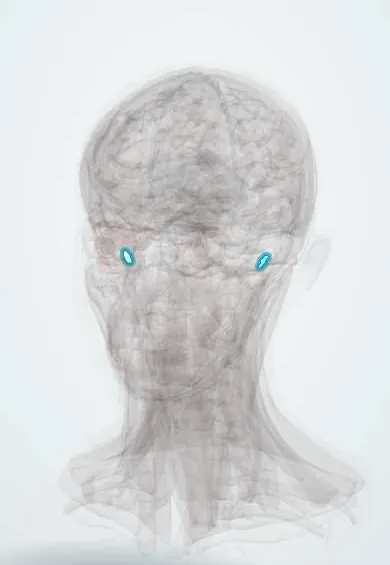
Representative model, for educational purposes only.