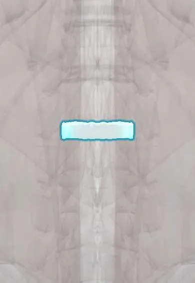
L1–L2 disc !
The L1-L2 disc is the cushion between the L1 and L2 vertebrae in the lower back; it absorbs shock and lets the spine bend and twist.
Official name: L1-L2
Opens in the Durgical app, no sign-up needed.
What it is
The cushion between L1 and L2: a tough outer ring of fibres (annulus fibrosus) around a soft centre.
What it does
Absorbs shock, spreads load across the vertebrae and lets the spine bend and twist, while holding the bones together.
Key facts
- Where
- Between L1 and L2 bodies; thick lumbar disc, taller in front (lumbar lordosis)
- Attaches
- Annulus fibrosus anchored to the endplates and ring apophyses of L1 and L2; anterior and posterior longitudinal ligaments
- Function
- Absorbs and spreads load; posterolateral herniation usually compresses the L2 root
Relations
- In front
- Abdominal aorta on the left and inferior vena cava on the right; the right and left crura of the diaphragm attach to the bodies at this height
- Behind
- Posterior longitudinal ligament, the dural sac, and the conus medullaris with the start of the cauda equina
- Lateral
- Psoas major arising from the sides of the bodies and disc, with the lumbar plexus forming inside it; the sympathetic trunk runs along the medial edge of the psoas major
How to find it
Several levels above the Tuffier line (the line joining the tops of the iliac crests, about L4). On X-ray it is found by counting down from T12, the last vertebra with a rib, or up from the sacrum.
Examination
- Femoral nerve stretch test (prone, knee bent, hip extended): pain in the front of the thigh suggests irritation of the upper lumbar roots (L2–L4)
- Hip flexion against resistance with the person seated: weakness points to the L2 root
On imaging
Radiographs show only disc height and spurs. Sagittal MRI shows where the conus ends, often at about this level, and whether a herniation presses on it or on the cauda equina; axial images show which root is involved. Upper lumbar herniations are uncommon, so MRI also looks for other causes such as a tumour or infection (discitis: fluid in the disc, oedema in both endplates).
Common problems
- Herniation (slipped disc): the gel pushes through a tear in the ring and presses on a nerve root, causing sciatica down the leg.
- Degeneration with age: the disc dries out, loses height and can bulge, sometimes with back pain.
- Tears in the ring (annular fissures) that can cause back pain.
How it is diagnosed
History and exam (the straight-leg raise test, strength, reflexes and feeling in the leg). MRI shows the disc and which nerve is pressed; it is usually done only if pain lasts more than about 6 weeks or there are warning signs.
Treatment
- Most herniations improve within weeks to months: staying active, physical therapy, pain medicine, sometimes an epidural steroid injection.
- Severe or lasting leg pain, or weakness: microdiscectomy to remove the herniated piece.
- Urgent surgery if there are signs of cauda equina syndrome (numbness in the saddle area, bladder or bowel changes).
- Selected cases of disc collapse with instability: fusion with a cage and bone graft.
Clinical notes
- The thoracolumbar junction (T12–L2) is the commonest site of spinal fractures; burst and flexion-distraction injuries can disrupt this disc
- A lesion of the conus medullaris causes early bladder and bowel problems with saddle numbness, often with less leg pain than a cauda equina lesion
Development
The spinal cord stops lengthening early while the spine keeps growing, so its lower end rises in relation to the vertebrae: it lies lower in the newborn (around L2–L3) and reaches about L1–L2 in the adult. A tethered cord blocks this rise and keeps the conus low.
Test yourself
Think of the answer, then tap the question to see it.
What is the L2 root picture from an L1–L2 disc herniation?
Weak hip flexion (iliopsoas) and numbness of the groin and upper front of the thigh; there is no reliable reflex.
Why are lumbar puncture and spinal anaesthesia done below the L1–L2 disc?
The conus medullaris ends near L1–L2 in most adults, so the needle goes lower (usually L3–L4 or L4–L5) to stay clear of it.
Which test suggests irritation of the upper lumbar roots, as with an L1–L2 disc herniation?
The femoral nerve stretch test: prone, knee bent, hip extended; pain in the front of the thigh suggests L2–L4 root irritation.
Which muscle arises from the sides of the L1–L2 disc, and what forms inside it?
The psoas major; the lumbar plexus forms inside it, and the sympathetic trunk runs along its medial edge.
Educational information, not medical advice. For any symptom, see a health professional.
Its names
- Official Latin (TA2)
- discus intervertebralis L1-L2 TA2 1684
- Official Spanish (DeCS)
- Disco L1-L2 DeCS D007403
- English (TA2)
- L1-L2
- Also called
- fibrous ring · fibrocartilago intervertebralis
Related
Sources: Terminologia Anatomica (TA2); DeCS; Anatomy, Back, Intervertebral Discs; 13.2 The Central Nervous System (OpenStax (Rice University), 2022) · adapted, CC BY 4.0.
Spotted a mistake?We are always improving for the community. Tell us what is wrong and our team will review it.Report a mistake
Representative model, for educational purposes only.