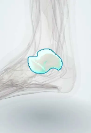
Clubfoot (talipes equinovarus) !
Clubfoot is a birth deformity in which the foot points down and turns inward; most cases have no known cause.
Updated 2026-10-29
Structures involved
Tap one to see what it is and open it in 3D.
- Talus
- Calcaneus
- Navicular
- Tibialis posterior
- Gastrocnemius (inner head)
- Soleus
- Cuboid
- Deltoid ligament
- Spring ligament
- Tibialis anterior
In embryology
- Developmental dysplasia of the hip
- Clubfoot (talipes equinovarus)
- Neural tube defects: anencephaly, spina bifida and encephalocele
- Malformation, disruption, deformation, sequence, syndrome, association
- Methotrexate and misoprostol
Why it happens
What it is
A congenital deformity in which the foot points down and turns inward, with a high arch, forefoot adduction, hindfoot varus and equinus. Most cases are idiopathic and present at birth.
Who gets it
One of the most common congenital musculoskeletal anomalies. It is more frequent in boys and is bilateral in about half of cases. Most affected children live in low- and middle-income countries, where the Ponseti method has been widely adopted.
Causes
- Idiopathic in most cases, with genetic and environmental contributions
- Syndromic or neuromuscular: myelomeningocele, arthrogryposis and other syndromes
- Positional (postural) deformity from crowding in the womb, which corrects easily
Risk factors
- Family history
- Male sex
- Maternal smoking in pregnancy
- Neural tube defects and neuromuscular disease
- Oligohydramnios
Pathophysiology
The talus, calcaneus and navicular are malpositioned: the navicular sits medially on the talar head and the calcaneus is turned under the talus. Medial and posterior soft tissues, including the tibialis posterior and the Achilles tendon, are short and tight, and the calf is thin. The deformity is described as cavus, adductus, varus and equinus (CAVE).
Classification
- Idiopathic, syndromic or neuromuscular, and positional
- Severity scores such as the Pirani and Dimeglio scores, which also track correction
- Atypical or complex clubfoot
Symptoms and signs
- Foot turned down and inward from birth
- Deep creases on the medial side and at the back of the heel
- High arch and curved lateral border
- Smaller foot and thinner calf on the affected side
- If untreated, walking on the outer border or top of the foot with painful calluses
Diagnosis
- Clinical
- Diagnosis is clinical at birth, separating a stiff clubfoot from a flexible positional foot. The examination includes the hips, spine and neurological system to find syndromic causes; prenatal ultrasound can detect it before birth.
- Tests
- Genetic testing only if a syndrome is suspected.
- Imaging
- Usually not needed for idiopathic clubfoot; X-rays or ultrasound in atypical cases or later follow-up; hip ultrasound if hip dysplasia is suspected.
Differential diagnosis
- Positional (postural) clubfoot
- Metatarsus adductus
- Congenital vertical talus
- Clubfoot from neuromuscular disease (myelomeningocele, arthrogryposis)
Goal of treatment
Achieve a plantigrade, flexible, painless foot that fits normal shoes, starting treatment in the first weeks of life and preventing relapse.
Treatment
- Conservative
- Ponseti method: gentle manipulation and weekly long-leg casts, correcting the cavus first, then adductus and varus together, and equinus last.
- Procedure
- Percutaneous Achilles tenotomy to correct remaining equinus, needed in most children treated with the Ponseti method.
- Conservative
- Foot abduction brace (boots and bar), full-time at first and then at night and naps for several years to prevent relapse.
- Surgical
- Tibialis anterior tendon transfer for dynamic relapse in older children; extensive soft-tissue release or bone surgery is reserved for resistant or neglected cases.
- Follow-up
- Regular follow-up through childhood to detect relapse early.
Complications
- Relapse, mostly linked to poor brace use
- Residual stiffness or under-correction
- Skin sores from casts or brace
- Stiffness, pain and arthritis after extensive surgical release
- Disability and difficulty walking if untreated
Prognosis
With the Ponseti method most children achieve a functional, painless foot and lead normal active lives. The foot and calf usually stay somewhat smaller.
Test yourself
Think of the answer, then tap the question to see it.
What four components make up clubfoot?
Cavus, forefoot adductus, hindfoot varus and equinus (CAVE).
In what order does the Ponseti method correct the deformity?
Cavus first, then adductus and varus together, and equinus last, usually with an Achilles tenotomy.
What is the main cause of relapse after Ponseti treatment?
Poor adherence to the foot abduction brace.
How is a positional foot told apart from a true clubfoot?
A positional foot is flexible and can be corrected passively to neutral; a true clubfoot is stiff.
Which conditions must be looked for in a newborn with clubfoot?
Neuromuscular and syndromic causes such as myelomeningocele or arthrogryposis, and other anomalies including hip dysplasia.
Related conditions
Educational information, not medical advice. For any symptom, see a health professional.
Sources: Talipes Equinovarus (StatPearls (NCBI Bookshelf), 2026).
Spotted a mistake?We are always improving for the community. Tell us what is wrong and our team will review it.Report a mistake
Representative model, for educational purposes only.