Shoulder dislocation !
A shoulder dislocation is when the ball of the upper arm bone slips out of its socket, usually forward; it is the large joint that dislocates most often.
Updated 2026-10-29
Structures involved
Tap one to see what it is and open it in 3D.
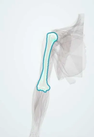
- Humerus
- Scapula
- Labrum
- Shoulder capsule
- Axillary nerve
- IGHL
- Humeral head cartilage
- Glenoid cartilage
- Axillary artery
- Supraspinatus
What it is
Loss of contact between the humeral head and the glenoid of the scapula. The glenohumeral joint is the most commonly dislocated large joint, and most dislocations are anterior.
Who gets it
Seen most often in young men after sport or falls and in older adults after falls. Recurrence is high in adolescents and young adults.
Causes
- Anterior: fall or blow with the arm abducted and externally rotated
- Posterior: seizures, electric shock or a direct blow from the front
- Inferior (luxatio erecta): forced hyperabduction, rare
Risk factors
- Contact and overhead sports
- Young age (for recurrence)
- Previous dislocation
- Ligamentous laxity
- Epilepsy (posterior dislocation)
- Falls in older adults
Pathophysiology
The shallow glenoid gives a wide range of motion but little bony stability, so the joint depends on the labrum, capsule and glenohumeral ligaments. In anterior dislocation the head is levered forward, often tearing the anteroinferior labrum (Bankart lesion) and impacting the back of the head on the glenoid rim (Hill-Sachs lesion). These lesions explain the frequent recurrence.
Classification
- Anterior (most common; subcoracoid is the usual type)
- Posterior
- Inferior (luxatio erecta)
- First-time or recurrent; traumatic or atraumatic
Symptoms and signs
- Severe shoulder pain after injury
- Arm held in slight abduction and external rotation (anterior)
- Arm held in internal rotation, unable to rotate outwards (posterior)
- Loss of the rounded shoulder contour, with a prominent acromion
- Numbness over the lateral shoulder if the axillary nerve is injured
Diagnosis
- Clinical
- Deformity and painful, restricted movement. Neurovascular examination before and after reduction, especially axillary nerve sensation over the lateral deltoid and distal pulses.
- Tests
- Laboratory tests are not needed for an isolated dislocation.
- Imaging
- X-ray before reduction (AP plus scapular Y or axillary view) to confirm direction and exclude fracture; repeat X-ray after reduction. MRI or MR arthrography shows labral (Bankart) and cuff injury; CT assesses glenoid bone loss and Hill-Sachs size.
Differential diagnosis
- Proximal humerus fracture
- Acromioclavicular joint separation
- Clavicle fracture
- Rotator cuff tear
- Posterior dislocation missed on a single AP view
Goal of treatment
Reduce the joint promptly and safely, protect nerves and vessels, and lower the risk of recurrent instability.
Treatment
- Acute
- Closed reduction with analgesia or procedural sedation, using one of several established techniques; neurovascular check before and after.
- Conservative
- Short period in a sling for comfort after reduction.
- Rehabilitation
- Physiotherapy to restore range of motion and strengthen the rotator cuff and scapular muscles.
- Surgical
- Stabilisation (arthroscopic Bankart repair or bone-block procedures for glenoid bone loss) for recurrent instability and, in some centres, after a first dislocation in young athletes; open reduction if closed reduction fails.
Complications
- Recurrent dislocation and chronic instability
- Axillary nerve injury
- Rotator cuff tear (more common over about 40)
- Greater tuberosity or glenoid rim fracture
- Axillary artery injury (rare)
- Later glenohumeral arthritis
Prognosis
Most first dislocations reduce without difficulty. Recurrence is the main problem: it is highest in teenagers and young adults and falls with age, while older patients more often have cuff tears or nerve injury.
Prevention
- Cuff and scapular strengthening for athletes
- Fall prevention in older adults
Test yourself
Think of the answer, then tap the question to see it.
What is the most common direction of shoulder dislocation?
Anterior (usually subcoracoid), by a wide margin.
Which nerve must be checked in a shoulder dislocation, and how?
The axillary nerve: sensation over the lateral shoulder (regimental badge area) and deltoid function, before and after reduction.
What are the Hill-Sachs and Bankart lesions?
Hill-Sachs: impaction defect on the posterolateral humeral head. Bankart: tear of the anteroinferior labrum, sometimes with a glenoid rim fragment.
Which patients have the highest risk of recurrent shoulder dislocation?
Adolescents and young adults, especially those active in contact or overhead sports.
Which shoulder dislocation is classically linked to seizures or electric shock?
Posterior dislocation; the arm is held in internal rotation and it is easily missed on a single AP X-ray.
Related conditions
- Rotator cuff tear
- Proximal humerus fracture
- Acromioclavicular joint separation
- Clavicle fracture
- Brachial plexus injury
- Elbow dislocation
- Axillary nerve injury
Educational information, not medical advice. For any symptom, see a health professional.
Sources: Anterior Glenohumeral Joint Dislocation (StatPearls (NCBI Bookshelf), 2023).
Spotted a mistake?We are always improving for the community. Tell us what is wrong and our team will review it.Report a mistake
Representative model, for educational purposes only.