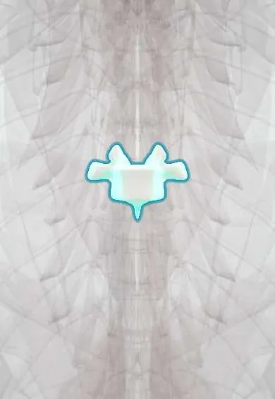
T8 vertebra !
T8 is the eighth thoracic (mid-back) vertebra; it has joints for the ribs and helps form the cage that protects the heart and lungs.
Official name: T8
Opens in the Durgical app, no sign-up needed.
What it is
The T8 mid-back vertebra, with joints on each side for the ribs.
What it does
Holds up the trunk and, with the ribs and breastbone, forms the rib cage, which protects the heart and lungs. The ribs make this part of the spine stiff.
Key facts
- Joints with
- T7 above and T9 below (discs, facet joints); heads of ribs 8 and 9; tubercle of rib 8 (costotransverse joint)
- Landmarks
- Typical thoracic: heart-shaped body; costal demifacets above and below; transverse costal facets; long sloping spine; caval opening of diaphragm (IVC) at T8
Relations
- In front
- Esophagus in front and slightly left of the descending thoracic aorta; the inferior vena cava pierces the diaphragm in front and to the right at this level
- Lateral
- Heads of the eighth and ninth ribs; the hemiazygos vein crosses to the azygos vein behind the aorta at about this level
- Behind
- Latissimus dorsi, lower trapezius and erector spinae
How to find it
One spine below T7, which is level with the inferior angle of the scapula; its tip lies over the body of T9.
On imaging
On a lateral view compression fractures are graded by height loss (Genant: mild about 20–25%, moderate 25–40%, severe over 40%). Fat-suppressed T2 or STIR MRI shows edema in a recent fracture; a bulging back wall, pedicle involvement or a soft tissue mass favour a tumor over osteoporosis.
Common problems
- Compression fractures from osteoporosis, sometimes after a minor fall or even a cough.
- A rounded upper back (kyphosis) from posture, wedge fractures or Scheuermann disease in teens.
- Scoliosis: a sideways curve with rotation, often starting in adolescence.
How it is diagnosed
X-rays for fractures and curves (scoliosis is measured with the Cobb angle). MRI tells new fractures from old ones. A bone density scan (DEXA) checks for osteoporosis.
Treatment
- Osteoporosis fractures: pain control, a short time in a brace, and treatment of the osteoporosis.
- Painful fractures that do not settle: kyphoplasty or vertebroplasty (cement injected into the bone).
- Large or progressive curves: bracing in growing children, sometimes fusion surgery.
Clinical notes
- Several mid-thoracic wedge fractures add up to a forward stoop (dowager's hump), loss of height and reduced lung capacity
- Wedge fractures high in the thoracic spine (above about T7; some texts T4) are unusual in osteoporosis and prompt a search for tumor or infection
- Vertebroplasty and kyphoplasty inject bone cement into painful fractures; leakage into the canal or the veins is the main risk
Memory aid
Diaphragm openings, 'I 8 10 Eggs At 12': Inferior vena cava at T8, Esophagus at T10, Aorta at T12.
Related conditions
Test yourself
Think of the answer, then tap the question to see it.
Which vessel passes through the diaphragm at the T8 vertebra, and why does its opening widen when the diaphragm contracts?
The inferior vena cava; it passes through the central tendon, so the opening widens rather than closes («I 8 10 Eggs At 12»).
In the Denis three-column model, what separates a compression fracture of a vertebra such as T8 from a burst fracture?
An intact back wall of the body: a compression fracture breaks only the anterior column.
Where do osteoporotic wedge fractures cluster in the spine?
In the mid-thoracic spine around T7–T8 and at the thoracolumbar junction (T12–L1).
Why does a wedge fracture high in the thoracic spine, above about T7, prompt further investigation?
Such fractures are unusual in osteoporosis, so a tumor or infection is looked for.
Educational information, not medical advice. For any symptom, see a health professional.
Its names
- Official Latin (TA2)
- vertebra thoracica VIII TA2 1059
- Official Spanish (DeCS)
- Vértebra dorsal D8 DeCS D013904
- English (TA2)
- T8
Related
- T7–T8 disc
- T8–T9 disc
- Posterior intercostal arteries
- Anterior longitudinal ligament
- Rib-to-spine ligaments
- Dura (dural sac)
- Endplates
- Mid-back facet joints
Sources: Terminologia Anatomica (TA2); DeCS; Anatomy, Back, Thoracic Vertebrae.
Spotted a mistake?We are always improving for the community. Tell us what is wrong and our team will review it.Report a mistake
Representative model, for educational purposes only.