Conditions · Hematology and oncology
Castleman disease !
A rare disorder of lymph nodes: one enlarged node in some people, or many nodes with fever and body-wide inflammation in others.
Updated 2026-11-01
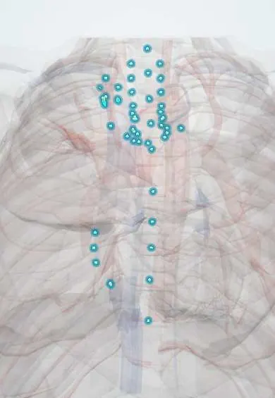
Structures involved
Tap one to see what it is and open it in 3D.
Why it happens
- Systemic effects of inflammation: fever, acute-phase proteins, leukocytosis, sepsis
- The physiology of inflammation
- Lymph node and spleen under the microscope
What it is
Rare lymphoproliferative disorders with a typical lymph node histology. The unicentric form affects one node or region (often mediastinum or neck) and is cured by surgery; the multicentric form causes IL-6-driven systemic inflammation and is either HHV-8-related (mostly with HIV) or idiopathic.
Who gets it
Rare; a few thousand new cases are estimated each year in the United States. Unicentric disease typically appears in young and middle-aged adults of either sex. HHV-8-associated multicentric disease occurs mostly in men with HIV, so it is relevant in Latin America, where HIV is often diagnosed late. Idiopathic multicentric disease, including the severe TAFRO form first described in Japan, occurs in middle-aged adults.
Causes
- Unicentric disease: a clonal proliferation of stromal (follicular dendritic) cells in many cases; cause otherwise unknown
- HHV-8-associated multicentric disease: the virus makes its own IL-6-like protein (viral IL-6) in infected plasmablasts
- Idiopathic multicentric disease: unknown; excess human IL-6 and other cytokines
- POEMS-associated multicentric disease: linked to a plasma cell clone
Risk factors
- HIV infection (HHV-8-associated form)
- Other immunosuppression with HHV-8 infection
- Plasma cell disorders (POEMS syndrome)
Pathophysiology
In multicentric disease, interleukin-6 and related cytokines produce fever, night sweats, weight loss, fatigue, anaemia, high CRP, low albumin, hypergammaglobulinaemia and fluid retention, and can progress to multiorgan failure. The nodes show regressed or hyperplastic germinal centres with abnormal vessels (hyaline-vascular pattern) or sheets of plasma cells between follicles (plasmacytic pattern). In unicentric disease, a large mass causes symptoms mainly by pressing on nearby structures.
Classification
- Unicentric Castleman disease: one node or region; mostly hyaline-vascular histology
- Multicentric, HHV-8-associated: usually with HIV; risk of Kaposi sarcoma and lymphoma
- Multicentric, idiopathic: iMCD-TAFRO (thrombocytopenia, anasarca, fever, reticulin fibrosis, renal failure, organomegaly) and iMCD-NOS
- Multicentric, POEMS-associated (polyneuropathy, organomegaly, endocrinopathy, monoclonal protein, skin changes)
Symptoms and signs
- Unicentric: often none; a mass found on imaging, or cough and chest discomfort from a mediastinal mass, or a neck lump
- Multicentric: generalised lymphadenopathy
- Fever, night sweats, weight loss and fatigue
- Enlarged liver and spleen
- Oedema, ascites and pleural effusions
- Rash, including cherry angiomas in idiopathic disease
- Numbness or weakness from neuropathy in POEMS
Diagnosis
- Clinical
- Consider it in a patient with an isolated mediastinal or neck mass, or with generalised lymphadenopathy, systemic inflammation and laboratory abnormalities that are not explained by infection, autoimmune disease or lymphoma. Glucocorticoids before biopsy can obscure the diagnosis.
- Tests
- Excisional lymph node biopsy (core or needle samples are usually insufficient) with HHV-8 staining (LANA-1). HIV test and HHV-8 viral load; blood count, CRP, albumin, immunoglobulins, kidney and liver function, IL-6 levels; serum protein electrophoresis and VEGF if POEMS is suspected. Diagnosis of idiopathic disease needs major and minor criteria and exclusion of mimics.
- Imaging
- CT of the neck, chest, abdomen and pelvis to count involved regions; PET-CT helps choose the most active node to biopsy.
Differential diagnosis
- Lymphoma (Hodgkin and non-Hodgkin)
- Tuberculous lymphadenitis
- HIV lymphadenopathy and other viral infections such as infectious mononucleosis
- Systemic lupus erythematosus and other autoimmune disease
- Sarcoidosis
- IgG4-related disease
Goal of treatment
Remove unicentric disease completely; in multicentric disease, block IL-6-driven inflammation or the HHV-8-infected B cells, and treat HIV.
Treatment
- Surgical
- Unicentric disease: complete surgical removal of the involved node or mass, which is usually curative; radiotherapy or other options if it cannot be removed.
- Medical
- Idiopathic multicentric disease: anti-IL-6 therapy (siltuximab, or the IL-6 receptor antibody tocilizumab) is first line, with corticosteroids in severe disease; other immunosuppressants or chemotherapy if there is no response.
- Medical
- HHV-8-associated multicentric disease: rituximab, with chemotherapy for severe disease or concurrent Kaposi sarcoma, plus antiretroviral therapy for HIV.
- Acute
- Severe flares such as TAFRO: intensive care support, high-dose corticosteroids and urgent anti-IL-6 or cytotoxic therapy.
- Follow-up
- Follow-up of symptoms, CRP, blood counts and albumin to detect relapse; nursing attention to fluid balance, daily weights and infection signs.
Complications
- Multiorgan failure in severe multicentric disease
- Kaposi sarcoma and lymphoma (HHV-8-associated disease)
- Infections from immunosuppression
- Paraneoplastic pemphigus and bronchiolitis obliterans (rare, unicentric disease)
- AA amyloidosis from chronic inflammation
Prognosis
Unicentric disease has an excellent prognosis after complete resection. Multicentric disease is chronic and can be fatal; outcomes have improved with anti-IL-6 therapy for idiopathic disease and with rituximab and antiretroviral therapy for HHV-8-associated disease.
Test yourself
Think of the answer, then tap the question to see it.
What is the treatment of unicentric Castleman disease?
Complete surgical removal of the involved lymph node, which is usually curative.
Which cytokine drives the symptoms of multicentric Castleman disease?
Interleukin-6.
Which virus causes multicentric Castleman disease in people with HIV?
Human herpesvirus 8 (HHV-8).
What does TAFRO stand for in idiopathic multicentric Castleman disease?
Thrombocytopenia, anasarca, fever, reticulin fibrosis, renal dysfunction and organomegaly.
Why is an excisional biopsy preferred for diagnosing Castleman disease?
The diagnosis depends on overall node architecture, which needle samples rarely show.
Related conditions
- HIV infection and AIDS
- Kaposi sarcoma
- Non-Hodgkin lymphoma
- Hodgkin lymphoma
- Cervical lymphadenopathy
- Tuberculosis
- Sarcoidosis
- Systemic lupus erythematosus
- Infectious mononucleosis
- Amyloidosis
- Peripheral neuropathy (diabetic and other)
Educational information, not medical advice. For any symptom, see a health professional.
Sources: Castleman Disease (National Organization for Rare Disorders (NORD), 2025).
Spotted a mistake?We are always improving for the community. Tell us what is wrong and our team will review it.Report a mistake
Representative model, for educational purposes only.