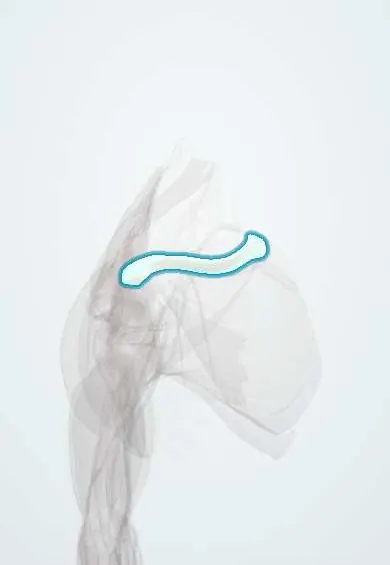
Clavicle fracture !
A clavicle fracture is a break of the collarbone, usually in its middle third; it is one of the most common fractures and most heal without surgery.
Updated 2026-10-29
Structures involved
Tap one to see what it is and open it in 3D.
What it is
Break of the collarbone, one of the most common fractures at all ages, usually in its middle third. Most heal without surgery, but the nearby subclavian vessels, brachial plexus and lung must be checked.
Who gets it
Common in children, adolescents and young adults, especially men, from falls, contact sports, cycling and motorcycle accidents; also seen in newborns as a birth injury and in older adults after simple falls.
Causes
- Fall onto the side of the shoulder (most common)
- Fall on an outstretched hand
- Direct blow to the clavicle
- Birth trauma in newborns (for example shoulder dystocia)
Risk factors
- Contact sports and cycling
- Motorcycle and road traffic accidents
- Young male sex
- Osteoporosis and falls in older adults
- Large birth weight or difficult delivery in newborns
Pathophysiology
The middle third is the thinnest part and lacks ligament and muscle support, so it breaks most often. After a midshaft fracture the medial fragment is pulled up by the sternocleidomastoid and the lateral fragment falls with the weight of the arm and is pulled medially, causing shortening. The subclavian vessels and brachial plexus lie just beneath.
Classification
- By location (Allman): middle third (most common), lateral (distal) third, medial third (rare)
- Lateral third fractures further divided by the integrity of the coracoclavicular (conoid and trapezoid) ligaments
- Non-displaced, displaced, shortened or comminuted
Symptoms and signs
- Pain over the clavicle after a fall or blow
- Visible or palpable deformity or bump
- Swelling and bruising
- Arm held against the body, supported by the other hand
- Painful or limited shoulder movement; crepitus
Diagnosis
- Clinical
- Local tenderness and deformity, with a full check of the skin, distal pulses, arm sensation and strength, and breath sounds.
- Tests
- Usually none; tests are added only for associated injuries in polytrauma.
- Imaging
- AP and cephalic-tilt X-rays of the clavicle confirm and measure displacement and shortening; CT for medial fractures or sternoclavicular involvement; chest X-ray if pneumothorax is suspected.
Differential diagnosis
- Acromioclavicular joint separation
- Sternoclavicular joint dislocation
- Proximal humerus fracture
- Shoulder dislocation
- Scapula fracture
Goal of treatment
Achieve painless union with acceptable alignment and full shoulder function, and detect early any injury to skin, vessels, nerves or lung.
Treatment
- Conservative
- Most fractures: a sling or figure-of-eight bandage for comfort, analgesia, and early gentle movement as pain allows.
- Surgical
- Open reduction and fixation with a plate or intramedullary device for open fractures, skin at risk, neurovascular injury or floating shoulder; often considered for marked shortening or displacement and some lateral fractures (criteria vary).
- Rehabilitation
- Progressive range-of-motion and strengthening exercises once pain settles, with gradual return to sport.
Complications
- Malunion with a visible bump or shortening
- Nonunion, more likely with displaced, shortened or lateral fractures
- Subclavian vessel or brachial plexus injury
- Pneumothorax
- Hardware irritation or infection after surgery
Prognosis
Most midshaft fractures heal well without surgery, especially in children, who remodel strongly. Displaced and shortened fractures in adults have higher rates of nonunion and symptomatic malunion.
Prevention
- Protective equipment in contact sports and cycling
- Road safety measures for motorcycle riders
- Fall prevention in older adults
Test yourself
Think of the answer, then tap the question to see it.
Which part of the clavicle is fractured most often?
The middle third (midshaft).
How are most clavicle fractures treated?
Non-operatively, with a sling for comfort, analgesia and early movement.
Which structures must be examined after a clavicle fracture because they lie just beneath it?
The subclavian artery and vein, the brachial plexus and the apex of the lung.
Why does the medial fragment of a midshaft clavicle fracture displace upward?
The sternocleidomastoid pulls it up, while the arm's weight pulls the lateral fragment down.
What is an absolute indication for surgery in a clavicle fracture?
An open fracture, skin at risk from tenting, or associated neurovascular injury.
Related conditions
- Acromioclavicular joint separation
- Proximal humerus fracture
- Shoulder dislocation
- Brachial plexus injury
- Radial head subluxation (pulled elbow)
Educational information, not medical advice. For any symptom, see a health professional.
Sources: Clavicle Fracture (StatPearls (NCBI Bookshelf), 2026).
Spotted a mistake?We are always improving for the community. Tell us what is wrong and our team will review it.Report a mistake
Representative model, for educational purposes only.