
Cryptogenic organizing pneumonia
Inflammation of the small airways and air sacs that looks like a pneumonia but does not improve with antibiotics. It usually responds well to corticosteroids.
Quick facts
- Key signs
- Flu-like illness · Cough · Breathlessness
- Diagnosis
- Transbronchial biopsy or cryobiopsy
Red flags ↓
No doses: the clinician sets them by the current guideline.
What it is
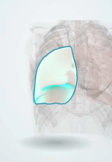
An idiopathic interstitial pneumonia in which plugs of loose granulation tissue fill the distal airways and alveoli. It is called cryptogenic when no cause is found; the same pattern secondary to infection, drugs, connective tissue disease, radiation or cancer is called secondary organizing pneumonia.
Who gets it
Uncommon, with an estimated few cases per 100 000 hospital admissions. It usually affects adults aged 50 to 60, men and women equally, and, unlike many interstitial diseases, it is more frequent in non-smokers.
Causes
- Cryptogenic: no cause found after a full search
- Secondary after infections: bacterial, viral (influenza, COVID-19) and others
- Drugs: amiodarone, nitrofurantoin, methotrexate, bleomycin, immune checkpoint inhibitors and others
- Connective tissue diseases (myositis, rheumatoid arthritis)
- Radiation therapy for breast cancer, transplantation, inhaled toxins and haematological cancers
Risk factors
- Age 50 to 60
- Recent respiratory infection
- Use of drugs known to cause organizing pneumonia
- Connective tissue disease
- Chest or breast radiotherapy
- Bone marrow or organ transplantation
Pathophysiology
Injury to the alveolar epithelium (cause unknown in the cryptogenic form; infection, drugs, radiation or rheumatic disease in secondary forms) → plasma proteins and fibrin leak into the alveoli → fibroblasts migrate into the exudate and organise it into buds of loose connective tissue (Masson bodies) in the alveoli, alveolar ducts and bronchioles → the air spaces fill, giving consolidation on imaging and impaired gas exchange → because the alveolar walls and architecture are preserved, the buds can be resorbed, so the process is largely reversible with corticosteroids.
Classification
- Cryptogenic versus secondary organizing pneumonia
- Typical (multiple patchy consolidations), focal (single mass-like lesion) and infiltrative (diffuse interstitial) imaging forms
- Acute fibrinous and organizing pneumonia: a severe variant with fibrin balls in the alveoli
Symptoms and signs
- Flu-like illness: fever, malaise, muscle aches
- Cough, usually dry
- Breathlessness of gradual onset over weeks to months
- Weight loss
- Inspiratory crackles over affected areas; clubbing is rare
- Symptoms that persist despite one or more courses of antibiotics
Diagnosis
- Clinical
- Suspected when a 'pneumonia' does not respond to antibiotics, with subacute fever, cough and breathlessness. A search for causes (drug history, infections, rheumatic disease, radiation, transplant) separates cryptogenic from secondary forms.
- Tests
- Raised inflammatory markers (ESR, C-reactive protein) and neutrophilia; negative cultures. Lung function shows a mild restrictive pattern with low DLCO. Bronchoalveolar lavage often has a mixed lymphocytosis with a low CD4/CD8 ratio and excludes infection. Transbronchial biopsy or cryobiopsy confirms organizing pneumonia; surgical biopsy if uncertain.
- Imaging
- Chest X-ray: bilateral patchy consolidations. CT: peripheral or peribronchial consolidations, often migratory (changing site), with air bronchograms and ground glass; the reversed halo (atoll) sign of central ground glass with a ring of consolidation suggests the diagnosis.
Differential diagnosis
- Community-acquired pneumonia and other infections (including tuberculosis)
- Chronic eosinophilic pneumonia
- Lung adenocarcinoma with lepidic growth or lymphoma
- Hypersensitivity pneumonitis
- Vasculitis such as granulomatosis with polyangiitis
- Pulmonary infarction
Consolidation that will not clear: organizing pneumonia or a mimic?
| Condition | Course | CT | Key test |
|---|---|---|---|
| Cryptogenic organizing pneumonia | Weeks of fever, cough and weight loss; no response to antibiotics | Patchy peripheral or peribronchial consolidation that migrates; reversed halo sign | Negative cultures; biopsy showing Masson bodies |
| Community-acquired pneumonia | Acute; improves within 48–72 hours of antibiotics | Lobar or segmental consolidation that stays in place | Response to antibiotics; cultures |
| Chronic eosinophilic pneumonia | Weeks of symptoms, often with asthma | Peripheral, upper-zone consolidation | Eosinophilia in blood and lavage |
| Lung adenocarcinoma or lymphoma | Slow, persistent, often without fever | Consolidation or ground glass that persists and never migrates | Biopsy |
| Tuberculosis | Weeks of cough, night sweats and weight loss; common in Colombia | Upper-lobe consolidation, cavities, tree-in-bud nodules | Sputum smear, molecular test and culture |
Goal of treatment
Confirm the diagnosis, remove any cause, and settle inflammation with corticosteroids while limiting their side effects and watching for relapse.
Treatment
- Medical
- Systemic corticosteroids are the mainstay, with a rapid response in days to weeks; they are tapered slowly over months because relapse is common when they are stopped early.
- Medical
- Steroid-sparing agents (for example macrolides in mild disease, or mycophenolate or azathioprine in relapsing disease) and stronger immunosuppression in fulminant cases.
- Follow-up
- Follow-up with symptoms, imaging and lung function to confirm resolution and catch relapse; bone protection, glucose checks and infection prophylaxis during long corticosteroid courses.
- Conservative
- Stop any causative drug and treat the underlying disease in secondary organizing pneumonia; mild, improving cases may be observed without treatment.
Complications
- Relapse (in up to a third of patients) during or after tapering
- Respiratory failure in fulminant or acute fibrinous forms
- Residual fibrosis in a minority
- Corticosteroid side effects: diabetes, osteoporosis, infections, weight gain
Common traps
- A pneumonia that fails antibiotics needs a stronger antibiotic: persistent or migrating consolidation with negative cultures should raise organizing pneumonia, and also tuberculosis and cancer
- Organizing pneumonia is a diagnosis in itself: it is a pattern; call it cryptogenic only after drugs, infection, connective tissue disease, radiation and cancer are excluded
- The reversed halo sign proves organizing pneumonia: it suggests it, but invasive fungal infection, tuberculosis and vasculitis can produce it too
- Once the patient feels better, corticosteroids can stop: relapse is common with a quick taper; they are reduced slowly over months
- Organizing pneumonia scars the lung like IPF: the architecture is preserved, so most patients recover fully
Prognosis
Good: most patients recover fully with corticosteroids. Relapses usually respond to retreatment and do not worsen survival. Fibrotic or fulminant forms and secondary organizing pneumonia with a severe underlying disease have a worse outlook.
Test yourself
Think of the answer, then tap the question to see it.
What clinical story suggests cryptogenic organizing pneumonia?
A subacute 'pneumonia' with fever, cough and breathlessness that does not improve with antibiotics, with patchy peripheral migratory consolidations.
What is the CT sign of central ground glass surrounded by a ring of consolidation?
The reversed halo or atoll sign, suggestive of organizing pneumonia.
What are Masson bodies?
Buds of loose granulation tissue (fibroblasts in a connective matrix) filling the alveoli and bronchioles: the hallmark of organizing pneumonia.
Why are corticosteroids tapered slowly in organizing pneumonia?
Because relapse is common when they are reduced quickly or stopped early.
A 55-year-old has 6 weeks of fever and cough despite two antibiotic courses; cultures and TB tests are negative; CT shows peripheral consolidations that have moved, one with a reversed halo. Next?
Organizing pneumonia. Search for a cause (drugs, rheumatic disease), biopsy if the picture is not convincing, then corticosteroids tapered slowly to prevent relapse.
Connections
Educational information, not medical advice. For any symptom, see a health professional.
Sources: Cryptogenic Organizing Pneumonia (StatPearls (NCBI Bookshelf), 2022); Interstitial Lung Disease (StatPearls (NCBI Bookshelf), 2026).
Spotted a mistake?We are always improving for the community. Tell us what is wrong and our team will review it.Report a mistake
Representative model, for educational purposes only.