
Endovascular aneurysm repair (EVAR)
A fabric-covered stent passed up from the groin lines a swollen aorta from inside. It avoids open surgery, but the aneurysm stays and is watched for life.
What it is
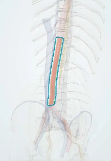
Repair of an abdominal aortic aneurysm with a stent graft delivered through the femoral arteries. The graft seals against healthy aorta above (the neck below the renal arteries) and healthy iliac arteries below, so blood flows through the graft and the aneurysm sac is excluded from pressure. Its typical complication is the endoleak: blood still reaching the sac.
Who gets it
EVAR has become a common way to repair abdominal aortic aneurysms because it has lower early mortality and a shorter stay than open repair. Long-term follow-up, however, shows more reinterventions, late ruptures and aneurysm-related deaths after EVAR, closely linked to endoleaks. Type II endoleak is the most common, seen in about 20–40% of patients after EVAR.
Causes
- Unruptured AAA that has reached the size threshold for repair, is growing fast or causes symptoms (thresholds and timing are on the abdominal aortic aneurysm page)
- Patients for whom open surgery is risky: anaesthetic risk, serious medical illness, or a hostile abdomen (scarring from earlier surgery, horseshoe kidney, stoma)
- Ruptured AAA with suitable anatomy: NICE notes EVAR benefits most people, especially men over 70 and women of any age, while open repair may suit men under 70 better
- Suitable anatomy: an adequate neck below the renal arteries (long enough, not too wide or angled), and iliac and femoral arteries that can carry the device and give a distal seal
Risk factors
- Short, wide, angled, calcified or thrombus-lined aortic neck (type Ia endoleak and migration)
- Short distal landing zone, a wide common iliac artery or tortuous iliacs (type Ib endoleak)
- Many patent lumbar arteries or a large patent inferior mesenteric artery (type II endoleak)
- Large aneurysm sac and ongoing sac growth
- Missed surveillance appointments
- Chronic kidney disease, which complicates contrast imaging
Pathophysiology
Once sealed, the sac is no longer pressurised and usually thromboses; in about half of patients it shrinks. Any route that refills it restores pressure on the old, weak wall. Leaks at the seals (type I) or through the graft (type III) carry full aortic pressure and risk rupture. Type II leaks run backwards from lumbar arteries, the inferior mesenteric artery or other side branches at lower pressure and often close on their own. As the sac remodels over the years, the graft can migrate, limbs can separate or kink and the seal can fail, so a well-sealed result early does not guarantee a well-sealed result later.
Classification
- Type I: failed seal at the proximal neck (Ia) or the iliac landing zone (Ib); high pressure, treated
- Type II: backflow into the sac from one (IIa) or several (IIb) side branches; low pressure, often resolves; treated if the sac grows
- Type III: flow through a junction between graft parts (IIIa) or a fabric tear (IIIb); high pressure, treated
- Type IV: flow through porous graft fabric in the first 30 days; resolves without treatment
- Type V (endotension): sac growth with no visible leak; investigated further
- Standard EVAR (infrarenal neck) versus complex EVAR (fenestrated or branched grafts for aneurysms reaching the renal arteries)
Symptoms and signs
- Usually none after EVAR: complications are found on surveillance imaging
- Groin pain, swelling or bruising at the access site
- Buttock or leg pain on walking, or a cold leg (a kinked or blocked graft limb)
- Fever and malaise, especially with abdominal or back pain (graft infection)
- Sudden abdominal or back pain with fainting (late rupture)
Diagnosis
- Clinical
- Before EVAR: confirm the indication for repair and assess fitness, life expectancy and the patient's ability to attend lifelong imaging, discussing EVAR, open repair and no repair. After EVAR: ask about leg claudication, groin problems and fever, and check that surveillance is up to date.
- Tests
- Kidney function before each contrast study, as contrast nephropathy and gadolinium restrictions shape the choice of scan; blood cultures and inflammatory markers when graft infection is suspected.
- Imaging
- CT angiography plans the repair (neck length, diameter and angle, iliac size and access). Surveillance is enrolled for life and its frequency is based on risk; one common pattern is imaging before discharge or at 1 month, at 6 and 12 months, then yearly. Colour duplex ultrasound or CT angiography follow sac diameter and limb kinking. If an endoleak is suspected, contrast CT angiography is used, or contrast-enhanced ultrasound when CT contrast is contraindicated; a negative colour duplex alone does not exclude an endoleak.
Differential diagnosis
- Open surgical repair (aneurysm replaced with a sewn-in graft)
- Conservative management with surveillance when the risks of repair outweigh rupture risk
- Complex (fenestrated or branched) EVAR when the aneurysm reaches the renal arteries
Endoleaks after EVAR: where the blood gets back in, and what is done
| Type | Source of flow | Pressure | Usual action |
|---|---|---|---|
| I (Ia, Ib) | Gap at the top (neck) or bottom (iliac) seal | High: full aortic pressure | Treat: balloon moulding, extension cuff or limb, or open conversion |
| II (IIa, IIb) | Backflow from lumbar arteries, inferior mesenteric artery or other side branches | Low | Watch; treat (often embolisation) if the sac grows |
| III (IIIa, IIIb) | Separation between graft parts or a tear in the fabric | High | Treat: reline with a new graft section |
| IV | Porous graft fabric, within 30 days | Low | No treatment; seals on its own |
| V (endotension) | Sac grows with no leak seen | Unclear; may hide a small leak | Investigate further; reline or open repair if growth continues |
Goal of treatment
Exclude the aneurysm from aortic pressure with less early risk than open surgery, then keep it excluded for life by detecting and fixing leaks, migration and limb problems before the sac ruptures.
Treatment
- Procedure
- Standard EVAR: a bifurcated stent graft is introduced through the femoral arteries under imaging and sealed against the infrarenal neck and the iliac arteries; the device is chosen to match the patient's anatomy.
- Procedure
- Type I and III endoleaks are repaired (open, endovascular or percutaneous): balloon moulding, a proximal cuff, extension into the external iliac artery, or relining with a new graft section.
- Procedure
- Type II endoleak is treated when the aneurysm sac grows: embolisation of the feeding branches or the sac (through the arteries, directly through the back or through the cava), or surgical ligation; recurrence after embolisation is common.
- Surgical
- Open conversion (removing the graft and repairing the aorta) for leaks or sac growth that cannot be controlled endovascularly, and for graft infection.
- Follow-up
- Lifelong surveillance imaging, more frequent than after open repair; patients are told plainly that EVAR needs follow-up for life.
- Medical
- Cardiovascular risk control as for all aneurysm patients: stopping smoking, blood pressure and lipid control and antiplatelet therapy when indicated.
Complications
- Endoleak (types I–V), the commonest reason for reintervention
- Graft migration, kinking or angulation, and separation of components
- Limb thrombosis with leg ischaemia
- Late rupture of the excluded sac
- Problems at the groin access site, such as bleeding and haematoma
- Contrast-related kidney injury from the procedure and repeated imaging
- Graft infection
- Radiation exposure from repeated CT, a concern in younger patients
Common traps
- After EVAR the aneurysm is gone: the sac stays in place and can be repressurised, so surveillance continues for life
- Every endoleak needs treatment: type II is low pressure and often closes on its own; it is treated when the sac grows, while types I and III are treated because they carry full aortic pressure
- A normal colour duplex scan excludes an endoleak: duplex misses some type I and III leaks; contrast CT angiography, or contrast-enhanced ultrasound, settles it
- EVAR is better than open repair because fewer patients die early: the early advantage fades, and reinterventions and late ruptures are more frequent, so younger fit patients and those unable to attend follow-up are often better served by open repair
- No leak on CT means the sac is safe even if it grows: sac growth without a visible leak (type V) is investigated, because a small leak may be hidden
Prognosis
Early survival is better than after open repair, but the advantage narrows over time because of reinterventions, endoleaks and late rupture. Outcomes depend heavily on suitable anatomy and on regular surveillance; a sac that shrinks and stays sealed carries a good outlook.
Prevention
- Careful CT planning and device choice that fits the neck and iliac anatomy
- Choosing EVAR only for patients who can attend lifelong surveillance
- Keeping surveillance up to date and acting on sac growth
- Stopping smoking and controlling cardiovascular risk
Test yourself
Think of the answer, then tap the question to see it.
Why does a patient need imaging for life after EVAR but not after open repair?
EVAR leaves the aneurysm sac in place. Endoleaks, migration or limb problems can repressurise it and cause late rupture, so the sac is checked regularly.
Which endoleak types are high pressure and usually treated?
Type I (failed seal at the neck or iliac landing zone) and type III (gap between graft parts or a fabric tear).
What is the most common endoleak, and when is it treated?
Type II, backflow from lumbar arteries or the inferior mesenteric artery. It often resolves and is treated when the sac keeps growing.
A duplex scan after EVAR shows no leak but the sac has grown. What next?
Look for a hidden leak with contrast CT angiography (or contrast-enhanced ultrasound); growth without a visible leak is type V and needs further work-up.
Which anatomical features make standard EVAR difficult?
A short, wide, angled, calcified or clot-lined neck below the renal arteries, and narrow or very tortuous iliac or femoral arteries.
Connections
Educational information, not medical advice. For any symptom, see a health professional.
Sources: Abdominal aortic aneurysm: diagnosis and management (NG156): Recommendations (National Institute for Health and Care Excellence (NICE), 2020); A Pictorial Review of Current Approaches in Endoleak Imaging (Interventional Radiology (Japanese Society of Interventional Radiology), 2025); Abdominal Aortic Aneurysms (AAA) (MSD Manual Professional Edition, 2025).
Spotted a mistake?We are always improving for the community. Tell us what is wrong and our team will review it.Report a mistake
Representative model, for educational purposes only.