
Acute aortic syndrome (intramural haematoma, penetrating aortic ulcer)
Three ways the aortic wall can fail suddenly: a tear (dissection), a bleed inside the wall, or an ulcer eating into it. They hurt alike and are treated alike.
What it is
An umbrella term for the acute, painful conditions of the aortic wall that threaten rupture: classic aortic dissection, intramural haematoma (bleeding within the media with no intimal tear seen on imaging) and penetrating atherosclerotic ulcer (a plaque ulcer that breaks through the internal elastic lamina into the media). They can coexist or turn into one another.
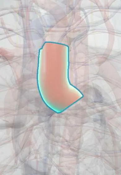
Who gets it
Classic dissection is the most common form; intramural haematoma and penetrating ulcer make up the rest, and about one in eight patients with a dissection also has one of them. Both are more frequent in older people with hypertension and atherosclerosis; penetrating ulcers mainly affect the descending thoracic aorta of elderly smokers.
Causes
- Intramural haematoma: bleeding from the vasa vasorum within the media, or a tiny intimal tear too small to see, usually on long-standing hypertension
- Penetrating atherosclerotic ulcer: an atherosclerotic plaque that ulcerates through the internal elastic lamina
- Classic dissection: an intimal tear in a weakened media (covered on the aortic dissection page)
- Trauma and iatrogenic injury (catheters, cardiac surgery) for all three
Risk factors
- Hypertension, the main factor for all three
- Older age, smoking and widespread atherosclerosis (especially for ulcers)
- An existing aortic aneurysm
- Connective tissue disorders and a bicuspid aortic valve (mostly classic dissection)
- Cocaine and other stimulants
Pathophysiology
A hypertensive, atherosclerotic or weakened wall → (1) the intima tears and blood splits the media (dissection), (2) a vessel inside the media bleeds with no flow channel (intramural haematoma), or (3) a plaque ulcer burrows through the internal elastic lamina (penetrating ulcer) → each thins and weakens the outer wall → expansion, rupture into the pericardium, pleura or mediastinum, or conversion: an intramural haematoma can develop a tear and become a dissection, or reabsorb; an ulcer can start a haematoma or a dissection, or form a false aneurysm. Involvement of the ascending aorta is the most lethal because of tamponade, aortic regurgitation and coronary involvement, and the mortality of untreated type A disease rises hour by hour.
Classification
- Stanford A: the ascending aorta is involved (any of the three lesions)
- Stanford B: the ascending aorta is spared
- 2024 ESC TEM classification: Type of syndrome, Entry tear location, Malperfusion
- Complicated (rupture, malperfusion, uncontrolled pain or blood pressure, rapid growth) versus uncomplicated
Symptoms and signs
- Sudden, severe chest or back pain, often worst at onset, as in dissection
- Pulse deficits and branch-vessel signs are less frequent than in classic dissection, because there is no flap to block the branches
- High blood pressure is common; low blood pressure suggests rupture or tamponade
Diagnosis
- Clinical
- Suspected in anyone with abrupt, severe chest, back or abdominal pain, especially with hypertension. A structured risk score (the aortic dissection detection risk score) combines high-risk conditions, pain features and examination findings.
- Tests
- ECG and troponin to look for coronary involvement and for the main alternative, an acute coronary syndrome. D-dimer can help rule out the diagnosis only in low-probability patients and may be less raised in intramural haematoma. Blood count, kidney function, lactate and cross-match.
- Imaging
- CT angiography of the whole aorta, cardiac-gated, with a non-contrast phase first. Intramural haematoma: a bright (hyperdense) crescent of smooth wall thickening on the non-contrast images that does not fill with contrast and has no flap. Penetrating ulcer: a contrast-filled crater projecting beyond the wall line among calcified plaques. Transoesophageal echocardiography in unstable patients and in theatre; MRI for follow-up.
Differential diagnosis
- Acute coronary syndrome
- Pulmonary embolism
- Pericarditis
- Symptomatic or ruptured aortic aneurysm
- Oesophageal rupture
- Musculoskeletal pain or renal colic
Acute aortic syndrome: the three lesions, and the one it is not
| Lesion | What happens in the wall | CT clue | Typical patient |
|---|---|---|---|
| Aortic dissection | Intimal tear; blood flows in a false lumen | Intimal flap with two lumens that fill with contrast | Hypertension; younger patients with connective tissue disease or a bicuspid valve |
| Intramural haematoma | Bleeding inside the media; no tear seen | Smooth crescent of wall thickening, bright before contrast, with no flow and no flap | Older, hypertensive |
| Penetrating atherosclerotic ulcer | A plaque ulcer breaks through the internal elastic lamina | Contrast-filled crater beyond the wall line among calcified plaques, often with haematoma | Elderly smokers with widespread atherosclerosis; descending aorta |
| Thoracic aortic aneurysm (not acute) | Slow widening of all wall layers | Dilated aorta with an intact wall, often with mural thrombus | Often painless until it expands or ruptures |
Goal of treatment
Prevent rupture and progression: control pain, heart rate and blood pressure at once, repair the ascending aorta urgently, and repair the descending aorta when the lesion is complicated.
Treatment
- Acute
- Intravenous analgesia and anti-impulse therapy: a beta-blocker first to slow the heart, then a vasodilator if blood pressure stays high, to targets set by the guideline.
- Surgical
- Stanford A dissection and intramural haematoma: emergency surgery in most patients; selected uncomplicated type A intramural haematomas in very high-risk patients may be managed medically with close imaging, as decided by an aortic team.
- Procedure
- Stanford B with complications (rupture, malperfusion, persistent pain, rapid growth, or a large or growing ulcer): thoracic endovascular aortic repair (TEVAR), or open surgery when the anatomy does not allow it.
- Follow-up
- Uncomplicated type B: intensive care monitoring, blood pressure control and repeat CT or MRI, because haematomas and ulcers can progress or regress.
- Medical
- Long term: blood pressure control, a statin and stopping smoking, with lifelong imaging follow-up of the aorta.
Complications
- Rupture into the pericardium (tamponade), pleura or mediastinum
- Progression of an intramural haematoma or ulcer to classic dissection
- Aneurysm or false aneurysm formation
- Aortic regurgitation and coronary involvement (type A)
- Organ or limb malperfusion
- Stroke and spinal cord ischaemia (paraplegia)
Common traps
- No flap on CT means no aortic emergency: an intramural haematoma has no flap and no flow; look at the non-contrast images for a bright crescent in the wall
- An intramural haematoma is a mild bruise that can wait: in the ascending aorta it behaves like a type A dissection and usually needs surgery
- A penetrating ulcer is just an ulcerated plaque: an ordinary ulcerated plaque stays within the intima; a penetrating ulcer crosses the internal elastic lamina into the media and can rupture
- Stanford applies only to dissection: the same rule, ascending aorta involved or not, guides treatment of haematoma and ulcer too
- A normal D-dimer excludes the syndrome: it helps only in low-probability patients and may be less raised in intramural haematoma; when suspicion is high, go straight to CT
Prognosis
Untreated type A lesions carry a high early mortality that rises hour by hour. Intramural haematomas can resolve, persist, or progress to dissection or aneurysm; ulcers that cause pain, are deep or enlarge carry a high risk of rupture. Survivors need lifelong blood pressure control and aortic imaging.
Prevention
- Blood pressure control and stopping smoking
- Surveillance and timely repair of aortic aneurysms
- Screening and follow-up of people with heritable aortic disease or a bicuspid aortic valve
Test yourself
Think of the answer, then tap the question to see it.
Which three lesions make up the acute aortic syndrome?
Classic aortic dissection, intramural haematoma (bleeding in the media without a visible intimal tear) and penetrating atherosclerotic ulcer (a plaque ulcer through the internal elastic lamina).
Why should CT for a suspected acute aortic syndrome include a non-contrast phase?
An intramural haematoma shows as a bright crescent of wall thickening before contrast; on contrast images alone it can be missed or mistaken for thrombus or wall thickening.
Which single anatomical question guides treatment of all three lesions?
Whether the ascending aorta is involved (Stanford A: usually emergency surgery) or not (Stanford B: medical control, with endovascular repair if complicated).
A 74-year-old hypertensive smoker has sudden severe back pain. CT: a contrast-filled crater in the descending aorta among calcified plaques, with wall haematoma. Pain persists. What next?
A symptomatic penetrating atherosclerotic ulcer (Stanford B) with persistent pain, so it is complicated: control heart rate and blood pressure and refer for endovascular repair (TEVAR).
Why it happens
Connections
Educational information, not medical advice. For any symptom, see a health professional.
Sources: Acute Aortic Syndrome (StatPearls (NCBI Bookshelf), 2024); Commentary on the 2024 European Society of Cardiology Guidelines on Peripheral Arterial and Aortic Diseases (European Cardiology Review, 2024); Aortic dissection: MedlinePlus Medical Encyclopedia (MedlinePlus, US National Library of Medicine, 2026).
Spotted a mistake?We are always improving for the community. Tell us what is wrong and our team will review it.Report a mistake
Representative model, for educational purposes only.