
Hip displacement in neuromuscular disease (cerebral palsy and spinal muscular atrophy)
In children with cerebral palsy or SMA who cannot walk, the hip often slips out slowly and painlessly. Regular X-rays catch it while it can be prevented.
What it is
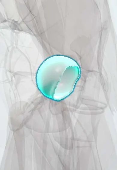
Neuromuscular hip displacement is the gradual lateral and upward migration of the femoral head out of the acetabulum in children with cerebral palsy, spinal muscular atrophy and similar conditions, from subluxation to complete dislocation. It is usually painless early, its risk rises as walking ability falls, and it is found by hip surveillance.
Who gets it
Hip displacement is the second most common musculoskeletal deformity in cerebral palsy, affecting about one third of children overall (35% in a large population study). Risk tracks the Gross Motor Function Classification System (GMFCS), not the motor type: it is very low at level I and about 90% at level V. It can begin as young as 2 years and progresses through childhood; in children who cannot walk the migration percentage rises by about 6–12% a year. In spinal muscular atrophy, displacement is common and starts earlier: about 84% at a mean age of 3 years in type 1 and 80% at about 6 years in type 2 in pooled reports.
Causes
- Cerebral palsy, especially spastic quadriplegia and GMFCS levels III to V
- Spinal muscular atrophy types 1 and 2 (weakness and low tone, not spasticity)
- Spina bifida (myelomeningocele)
- Other neuromuscular diseases and acquired brain or spinal cord injury in early childhood
Risk factors
- Not walking, or walking only with aids (GMFCS IV and V)
- Limited hip abduction from tight adductors and flexors
- Coxa valga, a raised head–shaft angle and increased femoral anteversion
- Pelvic obliquity, scoliosis and leg-length difference
- Positions held asymmetrically in sitting or lying
Pathophysiology
Weakness or spasticity prevents normal standing and walking → the hip abductors are weak relative to the adductors and flexors, and the femoral head is not loaded into the socket → the proximal femoral growth plate tilts and the femoral neck stays steep (coxa valga) with persistent anteversion → the femoral head drifts laterally and upward each year → the posterosuperior acetabulum fails to develop and becomes shallow → subluxation progresses to dislocation → cartilage of the femoral head is damaged, causing pain in many, plus problems with sitting, hygiene and pelvic obliquity with scoliosis. In spinal muscular atrophy the drive is weakness rather than spasticity.
Classification
- Hip at risk: migration percentage under 30%, monitored
- Displacement (subluxation): migration percentage of 30% or more, the usual threshold for orthopaedic referral
- Severe subluxation: over about 50%, when reconstructive surgery is usually recommended if the femoral head is still healthy
- Dislocation: migration percentage of 100%, the head fully out of the socket
Symptoms and signs
- Often no symptoms at first; found on surveillance radiographs
- Hip, groin or thigh pain, often shown as distress during changing, positioning or sitting
- Difficulty sitting upright, standing in a frame or tolerating seating
- Difficulty with perineal hygiene from tight adduction
- Signs: limited hip abduction, a hip flexion contracture, leg-length difference, pelvic obliquity; examination alone misses displacement
Diagnosis
- Clinical
- Hip surveillance combines a standard examination at each visit (hip abduction range, flexion contracture, leg length, pain, sitting posture, GMFCS level) with scheduled pelvic radiographs, because examination alone is unreliable. Surveillance continues at least to skeletal maturity, and a migration percentage of 30% or more, or hip pain, prompts referral to a paediatric orthopaedic surgeon.
- Tests
- No blood test diagnoses it. Bone health may be reviewed before surgery in children who do not walk.
- Imaging
- A standardised AP pelvis radiograph, starting by about 2 years in cerebral palsy and repeated at intervals set by GMFCS level and the migration percentage. The Reimers migration percentage is the key measure; the acetabular index and head–shaft angle add information. Arthrography or other imaging may help plan reconstruction.
Differential diagnosis
- Developmental dysplasia of the hip
- Traumatic hip dislocation
- Septic arthritis or osteomyelitis
- Femoral fracture in osteoporotic bone
- Other causes of pain in a child who cannot speak: dental, gastrointestinal or urinary problems
Hips that leave the socket: what drives them?
| Condition | Main driver | When it appears | Approach |
|---|---|---|---|
| Cerebral palsy | Spastic adductors and flexors with weak abductors; risk set by GMFCS | From about 2 years, through childhood | Surveillance by GMFCS level; soft-tissue release or reconstructive osteotomy |
| Spinal muscular atrophy | Weakness and low tone; coxa valga | Earlier: mean onset about 3 years in type 1 and 6 years in type 2 | Surveillance; individualised, from conservative care to proactive surgery |
| Developmental dysplasia of the hip | Shallow socket and lax capsule from birth | Newborn and infancy | Newborn screening; harness, reduction or surgery |
| Traumatic hip dislocation | High-energy injury | Any age, sudden | Emergency reduction |
Goal of treatment
Keep the hip comfortable and centred through growth so the child can sit, stand, be positioned and cared for without pain, while respecting the family's goals.
Treatment
- Follow-up
- Hip surveillance: scheduled examination and pelvic radiographs with migration percentage, more often at higher GMFCS levels and when displacement is found, until skeletal maturity.
- Conservative
- Postural management: positioning in abduction in lying, sitting and standing (seating systems, standing frames, abduction orthoses) and physiotherapy; these support comfort and posture, but there is no substantive evidence that they prevent displacement.
- Medical
- Spasticity management in cerebral palsy, such as botulinum toxin injections to the hip adductors; combined with an abduction brace it delayed surgery in a large trial but did not prevent displacement.
- Surgical
- Early displacement in young children: soft-tissue release (adductor and iliopsoas lengthening), which slows or stops progression in some. Progressive displacement: reconstruction with a varus derotation osteotomy of the proximal femur, often with a pelvic osteotomy; recurrence is more common when done before about 6–8 years. Guided growth of the proximal femur is a less invasive option with promising early results.
- Surgical
- Painful hips that cannot be reconstructed: salvage procedures such as proximal femoral resection with soft-tissue interposition, valgus osteotomy with femoral head resection, arthrodesis or joint replacement, chosen for comfort and care.
Complications
- Chronic hip pain and degeneration of the femoral head
- Loss of sitting balance and ability to stand for transfers
- Pelvic obliquity and scoliosis
- Pressure injuries and difficulty with hygiene
- Complications of surgery, which are frequent in children who do not walk
- Redisplacement after surgery, especially in spinal muscular atrophy
Common traps
- No pain means a healthy hip: displacement is silent for years and is found only by surveillance radiographs.
- Hip risk depends on the type of cerebral palsy alone: it is predicted best by GMFCS level, rising from near zero at level I to about 90% at level V.
- Botulinum toxin and bracing prevent hip dislocation: they can help tone and comfort, but a large trial found they delayed surgery without preventing displacement; progressive hips need surgery.
- SMA hips behave like cerebral palsy hips: they are driven by weakness rather than spasticity, displace earlier, and redisplacement after surgery is a concern, so care is more individualised.
- A dislocated hip must always be put back: in older children with a painless hip that does not affect sitting, observation is reasonable; treatment follows pain and function.
Prognosis
Hip surveillance with timely surgery reduces late dislocation and salvage surgery; in southern Sweden, 20 years of surveillance brought dislocations to very low rates. Late dislocation is often painful (over 80% in some studies) and harder to treat. In spinal muscular atrophy, disease-modifying therapies have improved survival and motor function, and hip care is changing as more children sit and stand.
Prevention
- Hip surveillance from early childhood in cerebral palsy and other high-risk conditions
- Early referral when the migration percentage reaches 30% or hip pain appears
- Postural care with abduction positioning
- Early disease-modifying therapy in spinal muscular atrophy
Test yourself
Think of the answer, then tap the question to see it.
What best predicts the risk of hip displacement in cerebral palsy?
The GMFCS level: risk is near zero at level I and up to about 90% at level V.
What does the migration percentage measure?
The proportion of the femoral head width lying outside Perkin line on an AP pelvis radiograph.
A 5-year-old with spastic quadriplegic cerebral palsy (GMFCS V) has no pain; migration percentage rose from 25% to 45% in a year. What next?
Referral to paediatric orthopaedics for surgery to keep the hip, such as soft-tissue release or femoral varus derotation osteotomy with pelvic osteotomy.
Why do hips displace in spinal muscular atrophy despite low muscle tone?
Weak hip abductors and lack of standing leave the femoral neck steep (coxa valga) and the socket shallow, so the head drifts out.
Connections
Educational information, not medical advice. For any symptom, see a health professional.
Sources: Hip Surveillance and Management of Hip Displacement in Children with Cerebral Palsy: Clinical and Ethical Dilemmas. (Journal of Clinical Medicine, 2023); Hip displacement management in spinal muscular atrophy in the era of disease modifying therapies: a Delphi consensus study in the UK. (EClinicalMedicine, 2026); The influence of tone on proximal femoral and acetabular geometry in neuromuscular hip displacement: A comparison of cerebral palsy and spinal muscular atrophy. (Journal of Children's Orthopaedics, 2022); Hip displacement in children with cerebral palsy: surveillance to surgery - a current concepts review (SICOT-J, 2024).
Spotted a mistake?We are always improving for the community. Tell us what is wrong and our team will review it.Report a mistake
Representative model, for educational purposes only.