
Chronic hypoventilation in neuromuscular and chest-wall disease
Weak breathing muscles or a stiff chest let carbon dioxide build up, first in sleep. Night-time mask ventilation relieves symptoms and can prolong life.
What it is
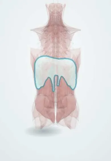
Chronic hypoventilation is a sustained rise in arterial carbon dioxide because the respiratory pump (nerves, breathing muscles or chest wall) cannot move enough air, while the lungs themselves may be normal. Causes include ALS, muscular dystrophies, spinal muscular atrophy, high spinal cord injury and severe kyphoscoliosis.
Who gets it
Chronic respiratory failure is a common complication of many neuromuscular diseases. In children the most frequent causes are Duchenne muscular dystrophy, spinal muscular atrophy and congenital muscle disorders; in adults, ALS, myotonic dystrophy and limb-girdle dystrophies. Speed varies: ALS and spinal muscular atrophy type 1 progress over months to a few years, Duchenne over years, some myopathies over decades.
Causes
- Motor neuron disease: amyotrophic lateral sclerosis, spinal muscular atrophy, post-polio weakness
- Muscle disease: Duchenne and limb-girdle muscular dystrophies, myotonic dystrophy, acid maltase deficiency (Pompe disease)
- Spinal cord and phrenic nerve: high cervical cord injury (C3–C5 supplies the diaphragm), bilateral phrenic nerve or diaphragm paralysis
- Neuromuscular junction and nerves, usually acute but can be chronic: myasthenia gravis, Guillain–Barré syndrome
- Chest wall: severe kyphoscoliosis, sequelae of tuberculosis surgery (thoracoplasty)
Risk factors
- Progressive neuromuscular disease, especially with bulbar weakness
- Weak cough, with repeated chest infections and secretion retention
- Coexisting obesity or obstructive sleep apnoea
- Sedatives or opioids that blunt breathing drive
- Unsupervised high-flow oxygen without ventilatory support
Pathophysiology
Weak inspiratory muscles produce small breaths; weak expiratory and bulbar muscles produce a poor cough, so secretions pool and atelectasis and infection follow. During REM sleep the accessory muscles lose tone and the diaphragm works alone, so a weak diaphragm first fails at night: CO2 rises and oxygen falls in REM. Repeated nocturnal hypercapnia resets the kidneys, which retain bicarbonate, and the respiratory centre tolerates a higher CO2; eventually hypercapnia persists by day. With bilateral diaphragm weakness, lying flat or bending forward lets the abdominal contents push the diaphragm up, so vital capacity falls further when supine.
Classification
- By site in the pump: drive, nerve, junction, muscle, chest wall
- By speed: rapidly progressive (ALS, SMA type 1), intermediate (Duchenne), slow (SMA type 3, many myopathies)
- By stage: nocturnal (REM) hypoventilation, then sleep-wide hypoventilation, then daytime hypercapnia
- Look-alikes: obesity hypoventilation (load on a normal pump), central hypoventilation (normal pump, low drive), COPD (lung disease)
Symptoms and signs
- Unrefreshing sleep, frequent awakenings, nightmares and nocturia
- Morning headache and daytime sleepiness
- Orthopnoea and breathlessness when lying flat, bending forward or in water (diaphragm weakness)
- Breathlessness when raising the arms or talking; short sentences
- Weak cough and repeated chest infections
- Often few complaints because the patient is too weak to exert
Diagnosis
- Clinical
- Ask about sleep quality, morning headache, orthopnoea and cough strength at every visit in neuromuscular disease, because symptoms appear late. Watch the patient breathe lying flat: inward movement of the abdomen during inspiration means a weak or paralysed diaphragm. Look for accessory muscle use, weak cough and scoliosis.
- Tests
- Spirometry upright and supine: a restrictive pattern, and a fall in vital capacity of about 30–50% when lying down points to bilateral diaphragm weakness. Respiratory muscle strength: maximal inspiratory pressure and sniff nasal pressure (usable with bulbar weakness); peak cough flow to judge cough. Overnight oximetry with transcutaneous or end-tidal CO2, or polysomnography, to detect REM hypoventilation. Arterial blood gas: raised PaCO2 with raised bicarbonate means chronic, compensated hypoventilation.
- Imaging
- Chest X-ray for elevated hemidiaphragms, atelectasis, infection and chest-wall deformity. Diaphragm ultrasound (excursion and thickening) or fluoroscopy with a sniff test performed supine shows absent or paradoxical movement.
Differential diagnosis
- Obesity hypoventilation syndrome
- Central hypoventilation (brainstem disease, opioids, hypothyroidism)
- COPD with chronic hypercapnia
- Obstructive sleep apnoea alone (normal daytime CO2)
- Heart failure (orthopnoea with oedema and crackles)
- Depression or poor sleep from other causes (headache and fatigue)
Hypercapnia with a normal lung: which pump is failing?
| Feature | Neuromuscular or chest wall | Obesity hypoventilation | COPD |
|---|---|---|---|
| Main problem | Weak muscles or stiff, deformed chest | Heavy chest and abdomen load a normal pump | Airflow obstruction and hyperinflation |
| Spirometry | Restrictive; vital capacity falls further supine | Mildly restrictive from body mass | Obstructive (low FEV1/FVC) |
| Clues | Weak cough, orthopnoea, paradoxical abdomen | Obesity, snoring, apnoeas, leg oedema | Smoking, wheeze, chronic cough |
| Night-time pattern | Hypoventilation worst in REM | Obstructive apnoeas plus hypoventilation | Desaturation in REM, often less CO2 rise |
| Main treatment | Nocturnal NIV plus cough assistance | NIV or CPAP plus weight loss | Bronchodilators; NIV in selected hypercapnic cases |
Goal of treatment
Rest the failing pump at night, keep the airway clear, prevent and treat chest infections early, and respect the patient's wishes about long-term ventilation.
Treatment
- Procedure
- Nocturnal non-invasive ventilation (usually bilevel pressure by nasal or full-face mask), started when symptoms of hypoventilation appear or tests show daytime hypercapnia, nocturnal hypoventilation, or low vital capacity or inspiratory pressure; it is extended into the day, sometimes by mouthpiece, as weakness progresses.
- Procedure
- Cough assistance: mechanical insufflation–exsufflation or manual techniques when peak cough flow is low, with early intensified airway clearance during chest infections.
- Medical
- Treat the cause where possible (for example disease-specific therapies in spinal muscular atrophy or Pompe disease, immunotherapy in myasthenia gravis) and treat chest infections promptly.
- Follow-up
- Regular review of symptoms, vital capacity, respiratory pressures, cough flow and overnight gas exchange; nurses check mask fit, skin pressure areas and secretions. Oxygen is added only with ventilatory support, because oxygen alone can mask or worsen hypercapnia.
- Conservative
- Advance care planning before a crisis: preferences about tracheostomy ventilation, hospital care and stopping ventilation, especially in rapidly progressive disease such as ALS.
Complications
- Acute-on-chronic hypercapnic respiratory failure, often triggered by a chest infection
- Pneumonia and atelectasis from retained secretions
- Pulmonary hypertension and right heart failure (cor pulmonale)
- Poor sleep, cognitive slowing and reduced quality of life
- Mask problems: nasal bridge pressure sores, air leaks, dry mouth
Common traps
- Normal daytime blood gases rule out hypoventilation: it starts in REM sleep, so ask about sleep and morning headache and test overnight.
- Low oxygen in a weak patient just needs more oxygen: the problem is ventilation, and oxygen alone can let CO2 climb further; support the breathing.
- Upright spirometry is enough: diaphragm weakness shows when the patient lies flat, so measure vital capacity supine as well.
- Few complaints mean the breathing is fine: patients who cannot walk do not exert themselves, so breathlessness is a late sign.
- Every hypercapnic obese patient has obesity hypoventilation: look for weak muscles, orthopnoea and a paradoxical abdomen, because neuromuscular disease can coexist.
Prognosis
Depends on the disease and its speed. In ALS non-invasive ventilation improves symptoms and quality of life and may prolong survival; in Duchenne muscular dystrophy ventilation and cough assistance have clearly extended life expectancy. In stable chest-wall disease nocturnal ventilation can keep patients well for many years.
Prevention
- Scheduled respiratory testing from diagnosis in neuromuscular disease
- Influenza and pneumococcal vaccination
- Early cough assistance before infections, and a written plan for chest infections
- Avoiding sedatives and unsupervised oxygen
Test yourself
Think of the answer, then tap the question to see it.
Why does hypoventilation from a weak diaphragm appear first during REM sleep?
In REM sleep the accessory and intercostal muscles lose tone, so breathing depends on the diaphragm alone; a weak diaphragm then cannot keep CO2 normal.
What spirometry finding suggests bilateral diaphragm weakness?
A restrictive pattern with a clear further fall in vital capacity when the patient lies supine compared with sitting.
Which bedside sign in the supine position suggests a paralysed diaphragm?
Paradoxical breathing: the abdomen moves inward during inspiration instead of outward.
Which test of respiratory muscle strength is useful when bulbar weakness prevents a good mouth seal?
Sniff nasal inspiratory pressure.
What is the main long-term treatment for chronic neuromuscular hypoventilation?
Nocturnal non-invasive ventilation, combined with cough assistance when cough is weak.
Connections
Educational information, not medical advice. For any symptom, see a health professional.
Sources: Noninvasive respiratory assistance as aid for respiratory care in neuromuscular disorders (Frontiers in Rehabilitation Sciences, 2023); Diaphragm dysfunction: how to diagnose and how to treat? (Breathe (Sheffield), 2025); Ventilatory Failure - Critical Care Medicine - MSD Manual Professional Edition (MSD Manual Professional Edition, 2025).
Spotted a mistake?We are always improving for the community. Tell us what is wrong and our team will review it.Report a mistake
Representative model, for educational purposes only.