
Hypertensive encephalopathy
A sudden, very high blood pressure overwhelms the brain's blood vessels and the brain swells: headache, confusion, seizures, blurred vision. It is an emergency.
What it is
Acute brain dysfunction caused by a rise in blood pressure severe enough to overcome the brain's control of its own blood flow, producing cerebral oedema. It is a form of hypertensive emergency and improves when the pressure is brought down in a controlled way.
Who gets it
Uncommon among people with hypertension. It is seen mostly with untreated or poorly controlled hypertension, stopped treatment, kidney disease and preeclampsia. People whose pressure was normal before can develop it at lower pressures than people with long-standing hypertension.
Causes
- Severe uncontrolled chronic hypertension, often after missed or stopped treatment
- Acute or chronic kidney disease
- Preeclampsia and eclampsia
- Secondary causes of hypertension
- Drugs that raise blood pressure, such as calcineurin inhibitors and some anti-cancer drugs
Risk factors
- Long-standing poorly controlled hypertension
- Stopping antihypertensive medicines
- Kidney disease
- Pregnancy with preeclampsia
- Immunosuppressive or anti-cancer drugs that raise blood pressure
Pathophysiology
Blood pressure rises abruptly → above the upper limit of autoregulation the cerebral arterioles can no longer constrict and are forced to dilate → flow surges and the high pressure reaches the capillaries → the blood–brain barrier leaks plasma into the brain (vasogenic oedema), most often in the posterior white matter, which has less sympathetic protection → headache, confusion, seizures and visual loss, with papilloedema and severe retinopathy in the eyes. In chronic hypertension the autoregulation range shifts to higher pressures: the brain tolerates more, but a sudden fall to 'normal' drops flow below the lower limit and causes ischaemia.
Classification
- One of the target-organ forms of hypertensive emergency (others: stroke, intracranial haemorrhage, acute heart failure, aortic dissection, acute kidney injury, eclampsia)
- Many cases show the imaging pattern of posterior reversible encephalopathy syndrome (PRES); hypertension is the commonest setting of PRES
Symptoms and signs
- Headache, often severe, with nausea or vomiting
- Confusion and drowsiness, progressing to stupor or coma
- Seizures
- Blurred vision or cortical blindness
- Global rather than focal deficits; focal signs, if present, are often brief and changing
Diagnosis
- Clinical
- Severe hypertension with global brain dysfunction (headache, confusion, seizures, visual change), usually with severe retinopathy or papilloedema on fundoscopy, that improves as the pressure is lowered. It is partly a diagnosis of exclusion: stroke, haemorrhage and other encephalopathies must be ruled out.
- Tests
- Urinalysis, electrolytes and creatinine for kidney damage; ECG for heart strain or ischaemia; a pregnancy test in women who could be pregnant.
- Imaging
- Head CT to exclude haemorrhage, infarct or a mass. MRI (FLAIR) shows vasogenic oedema, often posterior and on both sides, and diffusion imaging helps separate it from infarction.
Differential diagnosis
- Ischaemic stroke
- Intracerebral or subarachnoid haemorrhage
- Posterior reversible encephalopathy syndrome from a non-hypertensive trigger
- Eclampsia
- Encephalitis or meningitis
- Metabolic encephalopathy (uraemia, low sodium, low glucose)
- Post-ictal state
High pressure and a sick brain: which came first?
| Condition | Mental state and signs | Role of the blood pressure | Key imaging |
|---|---|---|---|
| Hypertensive encephalopathy | Global: headache, confusion, seizures, visual loss; few or changing focal signs | The cause: lowering it in a controlled way reverses the illness | Vasogenic oedema, often posterior; no infarct or bleed |
| Ischaemic stroke | Focal deficit in one arterial territory, often with normal alertness | Often a response; lowering it fast may harm the penumbra | Early CT often normal; diffusion MRI shows the infarct |
| Intracerebral haemorrhage | Sudden focal deficit, headache, vomiting, falling consciousness | Cause and consequence; lowered according to the guideline | CT shows bright blood |
| Eclampsia | Seizures in pregnancy or after delivery, with preeclampsia | Part of the disease; delivery is the definitive treatment | Often the PRES pattern |
Goal of treatment
Lower the blood pressure promptly but gradually, under close monitoring, to stop the brain swelling without dropping it so fast that a brain adapted to high pressure becomes ischaemic; then find and treat the cause.
Treatment
- Acute
- Intensive care with continuous blood pressure monitoring and a short-acting intravenous antihypertensive that can be adjusted minute to minute; the pressure is brought down progressively to guideline targets, not to normal at once. Oral drugs that drop the pressure unpredictably are avoided.
- Acute
- Seizure control with antiseizure medicines; magnesium sulfate when the cause is eclampsia.
- Follow-up
- Frequent neurological checks; if the patient does not improve as the pressure falls, repeat imaging and reconsider stroke, haemorrhage or another cause.
- Medical
- After recovery, long-term oral antihypertensive treatment, a search for secondary causes and support with adherence.
Complications
- Status epilepticus
- Cerebral infarction or haemorrhage, if untreated or if the pressure falls too fast
- Lasting visual loss
- Damage to other target organs: heart failure, kidney injury
- Coma and death
Common traps
- Bring the pressure to normal as fast as possible: a brain used to high pressure has a shifted autoregulation range, so a rapid fall causes ischaemia; the pressure is lowered gradually to guideline targets
- Very high pressure plus a neurological deficit means hypertensive encephalopathy: a focal deficit with normal alertness points first to stroke or haemorrhage, where the high pressure is often a response
- A normal head CT rules it out: CT can be normal or subtle; MRI shows the vasogenic oedema
- It only happens at extreme readings: people whose pressure was normal before, as in preeclampsia, can develop it at much lower values
- The diagnosis is certain on arrival: it is confirmed when the brain improves as the pressure comes down; if it does not, look for another cause
Prognosis
Usually fully reversible when the pressure is controlled promptly. Delay can lead to infarction, haemorrhage, coma or death.
Prevention
- Regular blood pressure checks and continuous treatment of hypertension
- Not stopping antihypertensive medicines abruptly
- Blood pressure monitoring in pregnancy
- Blood pressure monitoring in patients on drugs that raise it
Test yourself
Think of the answer, then tap the question to see it.
A man with untreated hypertension has a severe headache, confusion and blurred vision; fundoscopy shows papilloedema. What is the mechanism?
Hypertensive encephalopathy: pressure above the upper limit of autoregulation forces cerebral arterioles open, the blood–brain barrier leaks and vasogenic oedema develops.
Why is the blood pressure lowered gradually rather than straight to normal?
Chronic hypertension shifts autoregulation to higher pressures; a sudden fall drops cerebral blood flow below the lower limit and causes ischaemia.
Severe hypertension with right arm weakness in a fully alert patient: encephalopathy or stroke?
Stroke or haemorrhage first. Focal deficits with normal mental status suggest stroke; global dysfunction suggests encephalopathy. Brain imaging decides.
What does MRI typically show in hypertensive encephalopathy?
Vasogenic oedema on FLAIR, most often in the parieto-occipital white matter on both sides (the PRES pattern), without the restricted diffusion of an infarct.
What finding confirms the diagnosis in hindsight?
Clinical improvement as the blood pressure comes down; if the patient does not improve, another cause such as stroke, haemorrhage or infection must be sought.
Connections
Educational information, not medical advice. For any symptom, see a health professional.
Sources: Hypertensive Emergencies (MSD Manual Professional Edition, 2026); Posterior reversible encephalopathy syndrome (Journal of Neurology (open access, PMC), 2017).
Spotted a mistake?We are always improving for the community. Tell us what is wrong and our team will review it.Report a mistake
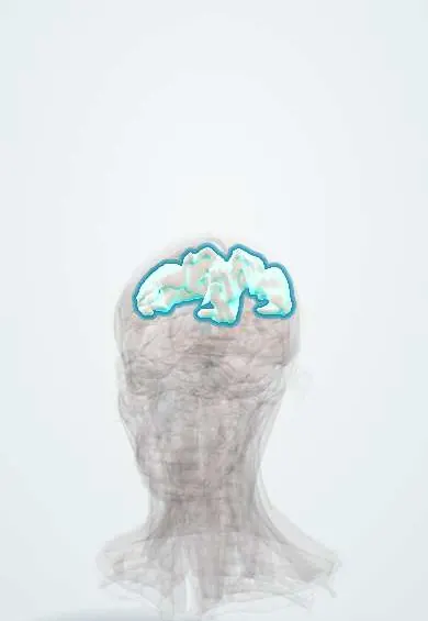
Representative model, for educational purposes only.