
Posterior reversible encephalopathy syndrome (PRES)
Swelling at the back of the brain, often from very high blood pressure, eclampsia or some drugs: headache, seizures, vision loss. It usually reverses.
What it is
An acute syndrome of headache, altered consciousness, seizures and visual disturbance with vasogenic brain oedema, typically in the parieto-occipital white matter on both sides. Triggers include severe hypertension, eclampsia, immunosuppressants, chemotherapy, kidney failure and sepsis; it usually reverses once treated.
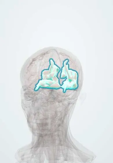
Who gets it
Reported at all ages, most often in young and middle-aged adults and more in women. It is found on MRI in most women with eclampsia and neurological symptoms, and in a few per cent of patients after organ or bone marrow transplantation. About 4 in 10 patients need intensive care for complications.
Causes
- Hypertension, often a sudden rise or a hypertensive emergency (most patients)
- Preeclampsia and eclampsia
- Immunosuppressants, especially calcineurin inhibitors such as tacrolimus, after organ, bone marrow or stem cell transplantation
- Cytotoxic chemotherapy and anti-angiogenic drugs (such as bevacizumab, sunitinib, sorafenib)
- Kidney failure (present in about half of patients)
- Sepsis
- Autoimmune disease, such as systemic lupus erythematosus
Risk factors
- High or swinging blood pressure
- Kidney disease
- Pregnancy with preeclampsia
- Transplantation and immunosuppressive treatment
- Cancer chemotherapy
- Autoimmune disease
- Low magnesium
Pathophysiology
Two routes, often combined, lead to the same leak. Pressure route: blood pressure rises above the upper limit of autoregulation → arterioles are forced open → hyperperfusion. Toxic route: circulating toxins or cytokines (eclampsia, sepsis, immunosuppressants, chemotherapy, autoimmune disease) injure and activate the endothelium → vasoconstriction and leaky junctions, even at modest pressures. Both → the blood–brain barrier lets plasma into the brain (vasogenic, not cytotoxic, oedema) → the posterior circulation, with less sympathetic innervation, is hit first → the subcortical white matter swells → headache, encephalopathy, seizures and visual loss. If the oedema is severe or the vessels clamp down, the tissue can infarct or bleed.
Classification
- Typical: bilateral parieto-occipital pattern (about 70% of patients)
- Variants: holohemispheric watershed and superior frontal sulcus patterns
- Atypical sites: brainstem, cerebellum, basal ganglia, spinal cord
- Complicated PRES: with haemorrhage (microbleeds or a haematoma), restricted diffusion (infarction) or contrast enhancement
Symptoms and signs
- Headache, nausea and vomiting
- Altered consciousness: confusion, somnolence, stupor or coma
- Seizures, focal or generalised (about two thirds of patients), sometimes status epilepticus
- Visual disturbance (about two thirds): blurred vision, field loss, cortical blindness, visual hallucinations
- Focal deficits in a minority
- Onset over hours to days, usually with high or swinging blood pressure
Diagnosis
- Clinical
- Suspected when acute neurological symptoms appear in a patient with a trigger (high or swinging pressure, eclampsia, transplant drugs, chemotherapy, kidney failure, sepsis, autoimmune disease). It is supported by the imaging pattern and confirmed, in hindsight, by reversal; other causes must be excluded.
- Tests
- No specific blood test. Kidney and liver function, magnesium and albumin (often low) and LDH; immunosuppressant levels (PRES can occur with levels in range); EEG for non-convulsive seizures; lumbar puncture when infection or leptomeningeal disease must be excluded (CSF may show raised protein without cells).
- Imaging
- MRI is the key: FLAIR and T2 show bright subcortical vasogenic oedema, usually symmetric and parieto-occipital; diffusion is usually not restricted, and restricted areas warn of infarction. Susceptibility-weighted sequences show microbleeds. CT is less sensitive. Angiography may show reversible vasoconstriction.
Differential diagnosis
- Posterior circulation ischaemic stroke (top of the basilar)
- Reversible cerebral vasoconstriction syndrome
- Cerebral venous thrombosis
- Encephalitis (infectious or autoimmune)
- Progressive multifocal leukoencephalopathy in immunosuppressed patients
- Osmotic demyelination or other demyelinating disease
- Metabolic or toxic encephalopathy
Bright posterior lesions: PRES or a look-alike?
| Condition | Setting | Clinical clue | Imaging clue |
|---|---|---|---|
| PRES | High or swinging pressure, eclampsia, transplant drugs, chemotherapy, kidney failure | Headache, seizures, visual loss, encephalopathy | Symmetric subcortical oedema without restricted diffusion; it reverses |
| Posterior circulation infarct (top of the basilar) | Vascular risk factors, embolism | Sudden onset, fixed deficits | Restricted diffusion within an arterial territory |
| Reversible cerebral vasoconstriction syndrome | After delivery or vasoactive drugs | Recurrent thunderclap headaches | Multifocal segmental narrowing on angiography; it can coexist with PRES |
| Progressive multifocal leukoencephalopathy | Severe immunosuppression | Deficits that worsen over weeks | Asymmetric white matter lesions that do not reverse |
Goal of treatment
Remove or treat the trigger, control the blood pressure without large swings and stop seizures; the brain usually recovers once the cause is gone.
Treatment
- Acute
- Controlled lowering of blood pressure as for a hypertensive emergency, with continuous, adjustable intravenous treatment under monitoring and without large swings.
- Acute
- Antiseizure treatment for seizures; it can usually be stopped once the patient is well and the lesions have resolved.
- Medical
- Stop, reduce or switch the triggering drug (calcineurin inhibitor, chemotherapy), deciding with the transplant or oncology team.
- Medical
- Treat the underlying condition: delivery and magnesium sulfate in eclampsia, treatment of sepsis, management of kidney failure, more immunosuppression when active lupus is the trigger.
- Follow-up
- Correct low magnesium; intensive care monitoring when consciousness falls or seizures persist; repeat MRI to confirm reversal.
Complications
- Status epilepticus
- Intracerebral or subarachnoid haemorrhage
- Cerebral infarction (areas of restricted diffusion)
- Raised intracranial pressure in severe cases
- Long-term epilepsy or persistent deficits in a minority
Common traps
- PRES is always posterior: frontal, watershed, brainstem and cerebellar patterns are common, and taking the name literally delays the diagnosis
- PRES is always reversible: most patients recover, but haemorrhage, infarction or status epilepticus can leave lasting damage
- PRES needs a very high blood pressure: about a third of patients have normal or only mildly raised pressure, especially with drug or toxic triggers
- A normal immunosuppressant level rules out drug-induced PRES: it occurs with levels in the therapeutic range, and reducing or switching the drug usually helps
- PRES oedema is cytotoxic like a stroke: it is vasogenic, so diffusion is usually not restricted; restricted diffusion suggests infarction and a worse outcome
Prognosis
Usually favourable: symptoms and imaging reverse in most patients once the trigger is treated. Outcome depends mainly on the underlying condition; a minority keep deficits or epilepsy, and haemorrhage, restricted diffusion or involvement of the corpus callosum predict a worse result.
Prevention
- Close blood pressure control in patients at risk (pregnancy, kidney disease, transplantation, chemotherapy)
- Monitoring blood pressure, magnesium and drug levels in patients on calcineurin inhibitors
- Prompt treatment of preeclampsia
Test yourself
Think of the answer, then tap the question to see it.
A kidney transplant recipient on tacrolimus has a headache, a seizure and blurred vision. MRI shows bright FLAIR signal in both occipital lobes. What is the likely diagnosis?
PRES triggered by the calcineurin inhibitor, often with raised blood pressure: control pressure and seizures and decide with the transplant team to reduce or switch the drug.
Why does PRES favour the back of the brain?
The posterior circulation has less sympathetic innervation, so it resists forced dilation less when pressure rises and leaks first.
What diffusion MRI finding separates PRES oedema from an infarct?
PRES is vasogenic oedema, usually without restricted diffusion; restricted diffusion means infarction and a worse prognosis.
Can PRES occur with a normal blood pressure?
Yes. About a third of patients have normal or mildly raised pressure, especially when a drug, sepsis or autoimmune disease injures the endothelium directly.
A week after delivery a woman has repeated thunderclap headaches, and angiography shows multifocal arterial narrowing. Which PRES look-alike is this?
Reversible cerebral vasoconstriction syndrome. It shares settings and imaging with PRES and can coexist with it; thunderclap headaches and segmental vasoconstriction point to it.
Connections
Educational information, not medical advice. For any symptom, see a health professional.
Sources: Posterior reversible encephalopathy syndrome (Journal of Neurology (open access, PMC), 2017); Posterior Reversible Encephalopathy Syndrome (PRES): Pathophysiology and Neuro-Imaging (Frontiers in Neurology (open access, PMC), 2020).
Spotted a mistake?We are always improving for the community. Tell us what is wrong and our team will review it.Report a mistake
Representative model, for educational purposes only.