Conditions · Infectious and tropical diseases
Intestinal amoebiasis (amoebic dysentery) !
Intestinal amoebiasis is a parasite infection of the colon from contaminated water or food; it can cause bloody diarrhoea and spread to the liver.
Updated 2026-11-01
Structures involved
Tap one to see what it is and open it in 3D.
Microorganisms that cause it
Why it happens
- Patterns of necrosis: coagulative, liquefactive, caseous, fat, fibrinoid, gangrenous
- Splanchnic and portal circulation
- Large intestine, appendix and anal canal
What it is
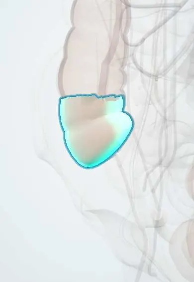
Infection of the large intestine by the protozoan Entamoeba histolytica, ranging from silent carriage to amoebic colitis with bloody diarrhoea (amoebic dysentery). It mainly affects the caecum and ascending colon and can spread to the liver.
Who gets it
Common in tropical regions with poor sanitation, including rural and peri-urban Colombia and much of Latin America. Most infections are asymptomatic. Microscopy cannot tell E. histolytica from harmless look-alike amoebae (E. dispar), so reported rates often overestimate true infection.
Causes
- Entamoeba histolytica, transmitted by ingesting cysts
- Water, food or raw vegetables contaminated with human faeces
- Direct faecal-oral contact, including oral-anal sex
Risk factors
- Living in or travelling to areas with poor sanitation and unsafe water
- Institutional settings and crowding
- Malnutrition, pregnancy and young age (more severe disease)
- Corticosteroid use or immunosuppression (fulminant colitis)
- Men who have sex with men
Pathophysiology
Swallowed cysts release trophozoites in the terminal ileum and colon. Trophozoites adhere to the mucosa, kill host cells and invade the submucosa, producing flask-shaped ulcers with normal mucosa between them, most often in the caecum and ascending colon. Trophozoites can enter the portal circulation and reach the liver to form an abscess.
Classification
- Asymptomatic intestinal infection (cyst passer)
- Non-dysenteric amoebic colitis
- Amoebic dysentery
- Fulminant necrotising colitis and toxic megacolon
- Amoeboma (inflammatory mass) and extraintestinal disease (liver abscess)
Symptoms and signs
- Gradual onset over 1–3 weeks of abdominal pain and cramps
- Diarrhoea with mucus and blood, often frequent and small in volume
- Tenesmus and right lower quadrant tenderness
- Weight loss and fatigue
- Fever in only a minority, unlike bacterial dysentery
Diagnosis
- Clinical
- Bloody diarrhoea with gradual onset, little fever and an exposure history. Corticosteroids should not be started for suspected inflammatory bowel disease until amoebiasis is excluded.
- Tests
- Stool antigen tests or PCR specific for E. histolytica (preferred); stool microscopy of several samples showing trophozoites with ingested red cells. Serology helps in invasive and liver disease. Full blood count, electrolytes and kidney function in severe cases.
- Imaging
- Colonoscopy or sigmoidoscopy shows discrete ulcers with normal mucosa between them; biopsy from the ulcer edge shows trophozoites. Abdominal X-ray or CT for megacolon or perforation; liver ultrasound when an abscess is suspected.
Differential diagnosis
- Bacterial dysentery (Shigella, Campylobacter, Salmonella, enteroinvasive E. coli)
- Ulcerative colitis and Crohn's disease
- Ischaemic colitis
- Colorectal cancer (amoeboma can mimic a tumour)
- Intestinal tuberculosis, Clostridioides difficile colitis
Goal of treatment
Kill invasive trophozoites, then eliminate cysts from the gut to prevent relapse and spread, correct dehydration, and detect complications early.
Treatment
- Medical
- A tissue amoebicide (a nitroimidazole such as metronidazole or tinidazole) for invasive disease.
- Medical
- Followed by a luminal agent (paromomycin, diloxanide furoate or iodoquinol) to clear cysts; asymptomatic E. histolytica carriers receive a luminal agent alone.
- Conservative
- Oral or intravenous rehydration and nutrition; antimotility drugs are avoided. Nursing care includes enteric precautions, hand hygiene and monitoring of fluid balance and stools.
- Surgical
- Laparotomy and colectomy for perforation, fulminant necrotising colitis or toxic megacolon.
Complications
- Amoebic liver abscess, the most common extraintestinal form
- Fulminant necrotising colitis and toxic megacolon
- Bowel perforation and peritonitis
- Amoeboma, strictures and intestinal obstruction
- Gastrointestinal bleeding and perianal skin ulceration
Prognosis
With a tissue amoebicide followed by a luminal agent most people recover in a few weeks. Fulminant colitis and perforation carry high mortality, and relapse is common if the luminal phase is skipped.
Prevention
- Safe drinking water (boiled, filtered or treated) and sanitation
- Handwashing after using the toilet and before handling food
- Washing or cooking vegetables and avoiding unpeeled raw fruit in high-risk areas
- Treating carriers, especially food handlers
Test yourself
Think of the answer, then tap the question to see it.
Which part of the colon is most often involved in amoebic colitis?
The caecum and ascending colon, followed by the rectosigmoid.
What is the classic endoscopic and histological lesion of amoebic colitis?
Discrete flask-shaped ulcers with normal mucosa between them; trophozoites with ingested red blood cells.
Why is a luminal agent given after a nitroimidazole?
Nitroimidazoles act on invading trophozoites but do not clear intestinal cysts; the luminal agent prevents relapse and transmission.
Why must amoebiasis be ruled out before giving corticosteroids for suspected ulcerative colitis?
Corticosteroids can turn amoebic colitis into fulminant necrotising colitis or toxic megacolon.
What is the most common extraintestinal complication of amoebiasis?
Amoebic liver abscess, usually in the right lobe, from spread through the portal vein.
Related conditions
- Amoebic liver abscess
- Acute (viral) gastroenteritis
- Giardiasis
- Typhoid (enteric) fever
- Ulcerative colitis
- Crohn disease
- Colorectal cancer
- Peritonitis
Educational information, not medical advice. For any symptom, see a health professional.
Sources: Amebiasis: MedlinePlus Medical Encyclopedia (MedlinePlus (U.S. National Library of Medicine), 2024); Amebiasis | Amebiasis | CDC (Centers for Disease Control and Prevention, 2024).
Spotted a mistake?We are always improving for the community. Tell us what is wrong and our team will review it.Report a mistake
Representative model, for educational purposes only.