Conditions · Infectious and tropical diseases
Paracoccidioidomycosis !
Paracoccidioidomycosis is a Latin American fungal infection of rural workers that scars the lungs and causes mouth ulcers and swollen neck nodes.
Updated 2026-11-01
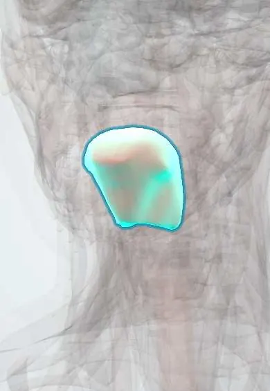
Structures involved
Tap one to see what it is and open it in 3D.
Microorganisms that cause it
Why it happens
- Granulomatous inflammation: caseating vs non-caseating, the causes to know
- Fibrosis in organs: TGF-β and the myofibroblast
- The alveolus: type I and II pneumocytes, surfactant, the blood–air barrier
- Oral cavity, tongue and teeth
What it is
A systemic endemic mycosis caused by fungi of the genus Paracoccidioides, acquired by inhaling spores from soil. It mainly affects the lungs, oral and upper airway mucosa, lymph nodes and adrenal glands. It is also called South American blastomycosis.
Who gets it
Restricted to Latin America, with the greatest burden in Brazil, followed by Colombia, Venezuela and Argentina. In Colombia it occurs mostly in humid coffee-growing and forested areas and affects mainly adult men who are farmers; the chronic form is far more common than the acute one.
Causes
- Paracoccidioides brasiliensis complex and P. lutzii, dimorphic fungi
- Inhalation of spores from soil during farm work
- Reactivation of latent infection years after exposure; no person-to-person spread
Risk factors
- Rural agricultural work in endemic areas, especially coffee and sugar cane
- Male sex (oestrogen inhibits the mould-to-yeast transition)
- Smoking and alcohol use
- Malnutrition and immunosuppression
Pathophysiology
Inhaled spores turn into large yeasts in the lungs. A strong cellular response forms granulomas that contain them, but the fungus can remain latent for decades. In the chronic adult form, reactivation causes progressive lung disease with fibrosis and emphysema, and spread to the oral, pharyngeal and laryngeal mucosa causes ulcers. In the acute or juvenile form, weak cellular immunity allows rapid spread to the lymphoid organs (lymph nodes, spleen, liver, bone marrow).
Classification
- Acute/subacute (juvenile) form: children and young adults, lymphoid organs
- Chronic (adult) form: unifocal or multifocal, lungs and mucosa
- Residual form: fibrotic sequelae
Symptoms and signs
- Chronic cough, sputum and breathlessness with relatively mild findings on exam
- Painful, granular oral ulcers with fine red dots ("mulberry-like" stomatitis), affecting gums, palate, lips and tongue
- Hoarseness and difficulty swallowing from laryngeal or pharyngeal involvement
- Large cervical lymph nodes that may suppurate and drain
- Weight loss, fever and malaise
Diagnosis
- Clinical
- Suspected in a rural worker from an endemic area with chronic cough, mouth ulcers and cervical nodes; the clinical picture often mimics tuberculosis or cancer.
- Tests
- Direct microscopy (KOH) of sputum, ulcer scrapings or node aspirate showing large yeasts with multiple buds ("pilot's wheel"); histopathology; culture (alert the laboratory: biohazard). Serology by double immunodiffusion helps diagnosis and follow-up. Check adrenal function and screen for coexisting tuberculosis.
- Imaging
- Chest X-ray or CT: bilateral, symmetric interstitial or nodular infiltrates in the middle and lower fields ("butterfly wings"), with emphysema and fibrosis.
Differential diagnosis
- Pulmonary tuberculosis (often coexists)
- Histoplasmosis
- Mucosal leishmaniasis
- Squamous cell carcinoma of the mouth or larynx
- Lymphoma
Goal of treatment
Control the infection with long antifungal therapy, prevent relapse, and limit lung and adrenal sequelae.
Treatment
- Medical
- Itraconazole is the drug of choice for mild to moderate disease, given for many months.
- Medical
- Trimethoprim-sulfamethoxazole as a cheaper alternative, often continued for more than two years.
- Acute
- Amphotericin B for severe or disseminated disease, then oral step-down therapy.
- Follow-up
- Clinical, radiological and serological criteria of cure before stopping; follow-up for relapse, adrenal function and lung function.
- Lifestyle
- Stopping smoking and alcohol and improving nutrition are part of management and reduce relapse.
Drugs for this condition
Complications
- Pulmonary fibrosis and emphysema with chronic respiratory failure
- Adrenal insufficiency
- Laryngeal and tracheal stenosis, microstomia
- Central nervous system involvement
- Relapse after stopping treatment
Prognosis
Treatment cures most patients, but the chronic form often leaves lung fibrosis; the acute juvenile form can be fatal if diagnosis is late.
Test yourself
Think of the answer, then tap the question to see it.
What is the classic microscopic finding of Paracoccidioides?
Large yeasts with multiple budding daughter cells, like a "pilot's wheel".
Who is typically affected by the chronic form?
Adult men who are rural agricultural workers, often smokers, in endemic Latin American areas.
Which endocrine gland is often involved?
The adrenal glands, causing adrenal insufficiency.
Which oral drug is first-line for mild to moderate disease?
Itraconazole (trimethoprim-sulfamethoxazole is an alternative).
Related conditions
- Histoplasmosis
- Pulmonary tuberculosis
- Leishmaniasis
- Laryngeal cancer
- Cervical lymphadenopathy
- Adrenal insufficiency
- Chronic obstructive pulmonary disease (COPD)
- Endemic mycoses: blastomycosis and coccidioidomycosis
- Hodgkin lymphoma
Educational information, not medical advice. For any symptom, see a health professional.
Sources: Paracoccidioidomycosis - Infectious Disease - MSD Manual Professional Edition (MSD Manual Professional Edition, 2024).
Spotted a mistake?We are always improving for the community. Tell us what is wrong and our team will review it.Report a mistake
Representative model, for educational purposes only.