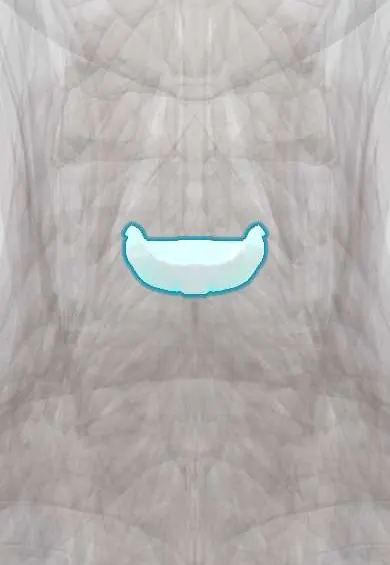
C4–C5 disc !
The C4-C5 disc is the cushion between the C4 and C5 neck vertebrae; it absorbs load and lets the neck bend and turn.
Official name: C4-C5
Opens in the Durgical app, no sign-up needed.
What it is
The cushion between C4 and C5: a tough outer ring of fibres (annulus fibrosus) around a soft centre.
What it does
Absorbs load and lets the neck bend and turn. C5-C6 is the most mobile and most worn neck level.
Key facts
- Where
- Between C4 and C5 bodies; taller in front (cervical lordosis); flanked by uncovertebral joints
- Attaches
- Annulus fibrosus anchored to the endplates and ring apophyses of C4 and C5; anterior and posterior longitudinal ligaments
- Function
- Absorbs and spreads load; posterolateral herniation usually compresses the C5 root
Relations
- In front
- Longus colli, then the esophagus and the larynx behind the thyroid cartilage; the carotid arteries and internal jugular vein lie anterolaterally in the carotid sheath
- Behind
- Posterior longitudinal ligament, the dural sac and the spinal cord at the start of its cervical enlargement
- Lateral
- Uncovertebral joints, the vertebral artery and the C5 nerve root in the C4–C5 foramen
How to find it
From the front, about the level of the thyroid cartilage (Adam's apple). As at every cervical level, the surgeon confirms it with a marker needle and a lateral X-ray before removing the disc.
Examination
- Shoulder abduction and elbow flexion against resistance: weakness of the deltoid and biceps points to the C5 nerve root
- Biceps reflex: reduced on the affected side with C5 (and C6) root compression
On imaging
Radiographs show narrowing and spurs; oblique views and CT show uncovertebral spurs encroaching on the C4–C5 foramen, where the C5 root exits. MRI is chosen for radiculopathy or myelopathy: axial T2 separates a soft disc in the foramen from bony narrowing, and sagittal images show cord compression. CT myelography is used when MRI is not possible.
Common problems
- Herniation pressing on the C6 nerve root: neck pain and pain, numbness or weakness down the arm into the thumb side of the hand.
- Degeneration with bone spurs, which can narrow the canal and press on the spinal cord (myelopathy: clumsy hands, unsteady walking).
How it is diagnosed
Exam of arm strength, reflexes and feeling, plus signs of cord pressure. MRI shows the disc, nerve roots and cord.
Treatment
- Most arm pain from a disc improves with time, physical therapy, medicine and sometimes an injection.
- Lasting arm pain, weakness or cord pressure: anterior cervical discectomy and fusion (ACDF), or disc replacement in selected patients.
Clinical notes
- Differentials for C5 weakness: rotator cuff tear, a brachial plexus stretch injury («burner» in contact sports) and neuralgic amyotrophy (Parsonage–Turner syndrome)
- A large central herniation at C4–C5 compresses the cord: lower motor neuron signs at the level (C5) with upper motor neuron signs below it, in arms and legs
Related conditions
Test yourself
Think of the answer, then tap the question to see it.
What is the clinical picture of the C5 root compressed by a C4–C5 disc herniation?
Weak shoulder abduction (deltoid) and elbow flexion (biceps), a reduced biceps reflex and numbness over the outer upper arm.
What is C5 palsy after cervical decompression?
Deltoid and biceps weakness appearing hours to days after surgery, often recovering over months.
How can C5 radiculopathy from a C4–C5 disc be told apart from a rotator cuff problem?
Pain that changes with neck movement rather than shoulder movement points to the nerve root.
Educational information, not medical advice. For any symptom, see a health professional.
Its names
- Official Latin (TA2)
- discus intervertebralis C4-C5 TA2 1684
- Official Spanish (DeCS)
- Disco C4-C5 DeCS D007403
- English (TA2)
- C4-C5
- Also called
- fibrous ring · fibrocartilago intervertebralis
Related
Sources: Terminologia Anatomica (TA2); DeCS; Anatomy, Back, Intervertebral Discs.
Spotted a mistake?We are always improving for the community. Tell us what is wrong and our team will review it.Report a mistake
Representative model, for educational purposes only.