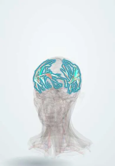
Primary angiitis of the central nervous system !
A rare inflammation of the blood vessels of the brain and spinal cord, with no disease elsewhere, that causes headache, confusion and repeated strokes.
Updated 2026-11-01
Structures involved
Tap one to see what it is and open it in 3D.
- Middle cerebral artery
- Circle of Willis
- Anterior cerebral artery
- Posterior cerebral artery
- Basilar artery
- Internal carotid artery
- White matter
- Temporal lobe
- Frontal lobe
Why it happens
- Infarction: red vs white, and what decides the damage
- Granulomatous inflammation: caseating vs non-caseating, the causes to know
- Cerebral blood flow, brain metabolism and intracranial pressure
- Blood vessels: artery, vein and capillary layers
What it is
An inflammatory disease of small and medium arteries confined to the brain, spinal cord and meninges, without vasculitis in other organs. Also called primary CNS vasculitis (PACNS). It typically produces a subacute picture of headache, cognitive decline and multifocal strokes, and is a diagnosis of exclusion that needs tissue or angiographic proof.
Who gets it
Very rare: roughly 2 to 3 new cases per million people per year. Median age at onset is around 50 years, with a slight male predominance. Secondary causes of CNS vasculitis (infections, systemic autoimmune disease, drugs) are far more common; in Colombia, syphilis, tuberculosis, varicella-zoster virus and HIV must be excluded first.
Causes
- Cause unknown; presumed autoimmune T-cell–mediated inflammation of cerebral vessel walls
- Some cases are linked to beta-amyloid deposits in vessel walls (amyloid-beta–related angiitis)
- Viral triggers such as varicella-zoster virus have been suspected but not proven in most cases
Risk factors
- Middle age (40 to 60 years)
- Male sex (slight predominance)
- Cerebral amyloid angiopathy in older adults (amyloid-beta–related form)
- No modifiable risk factors are known
Pathophysiology
Lymphocytes and macrophages infiltrate the walls of small and medium leptomeningeal and parenchymal arteries, sometimes forming granulomas or causing fibrinoid necrosis. The thickened, narrowed walls reduce flow and promote thrombosis, producing infarcts of different ages in several arterial territories, while inflammation of the meninges raises protein and cells in the cerebrospinal fluid. Lesions are patchy (skip lesions), which explains why a single biopsy can miss them.
Classification
- Granulomatous angiitis (most common histological pattern)
- Lymphocytic vasculitis
- Necrotising vasculitis
- Amyloid-beta–related angiitis (with cerebral amyloid angiopathy)
- Small-vessel (biopsy-proven, angiogram often normal) versus medium-vessel (angiogram abnormal) disease
Symptoms and signs
- Headache that is subacute, diffuse and progressive over weeks to months (most common symptom)
- Cognitive impairment, confusion or personality change (encephalopathy)
- Repeated strokes or transient ischaemic attacks in different territories
- Focal deficits: weakness, aphasia, ataxia or visual loss
- Seizures
- Spinal cord symptoms (paraparesis, sensory level) in a minority
Diagnosis
- Clinical
- Suspect it in a patient with unexplained, progressive neurological deficits plus headache and cognitive change. Diagnosis (Calabrese criteria) requires a new neurological deficit unexplained by other causes, plus either angiographic features of vasculitis or vasculitis on CNS biopsy, after systemic vasculitis and secondary causes have been excluded.
- Tests
- Lumbar puncture is abnormal in most cases (mild lymphocytic pleocytosis and raised protein) and is essential to exclude infection and lymphoma. Blood tests are usually normal (ESR and CRP often normal), which helps separate it from systemic vasculitis. Serology for syphilis, HIV, hepatitis, ANA, ANCA and antiphospholipid antibodies; varicella-zoster PCR and antibodies in CSF. Brain and leptomeningeal biopsy is the gold standard.
- Imaging
- MRI is almost always abnormal: multiple infarcts of different ages in several territories, white matter lesions and sometimes leptomeningeal enhancement; a normal MRI makes the diagnosis very unlikely. Catheter angiography may show beading (alternating narrowing and dilatation) of several arteries, but it is neither sensitive nor specific. Vessel wall MRI can show concentric wall thickening and enhancement.
Differential diagnosis
- Reversible cerebral vasoconstriction syndrome (thunderclap headache, normal CSF, reverses within weeks)
- Secondary CNS vasculitis from infection (varicella-zoster, syphilis, tuberculosis, HIV, fungi)
- Systemic lupus erythematosus and systemic vasculitis
- Cardioembolic stroke, including infective endocarditis
- Intravascular or primary CNS lymphoma
- Multiple sclerosis and other demyelinating disease
Goal of treatment
Induce remission quickly to prevent new infarcts, then maintain remission with less toxic immunosuppression while protecting the patient from infection, seizures and disability.
Treatment
- Acute
- Induction: high-dose glucocorticoids, usually starting intravenously, combined with cyclophosphamide (or mycophenolate or rituximab) in severe or rapidly progressive disease.
- Medical
- Maintenance: steroid-sparing immunosuppressants such as azathioprine, mycophenolate or methotrexate for one to two years or more, with slow tapering of glucocorticoids.
- Medical
- Supportive care: antiseizure drugs when there are seizures, antiplatelet therapy when large or medium vessels are involved, and Pneumocystis prophylaxis and bone protection during intense immunosuppression.
- Rehabilitation
- Neurorehabilitation (physiotherapy, occupational and speech therapy) for deficits after strokes.
- Follow-up
- Monitoring: serial neurological examination and MRI to judge response or relapse; nursing checks of glucose, blood pressure, infection signs and blood counts during treatment.
Complications
- Permanent neurological deficits and dementia
- Recurrent stroke or intracerebral haemorrhage
- Epilepsy
- Relapse after stopping treatment
- Opportunistic infections and other side effects of immunosuppression
Prognosis
Untreated disease was usually fatal; with immunosuppression most patients stabilise or improve, but around a quarter to a third relapse and many keep some disability. Large-vessel involvement, multiple infarcts and cognitive impairment at diagnosis predict a worse outcome.
Test yourself
Think of the answer, then tap the question to see it.
What is the gold standard for diagnosing primary angiitis of the CNS?
Biopsy of brain parenchyma and leptomeninges showing vasculitis.
How do CSF findings help separate PACNS from reversible cerebral vasoconstriction syndrome?
CSF is usually abnormal in PACNS (lymphocytes, raised protein) and usually normal in RCVS.
What headache pattern suggests RCVS rather than PACNS?
Recurrent sudden thunderclap headaches; PACNS usually causes a subacute, progressive headache.
Why are ESR and CRP of limited value in PACNS?
They are often normal because the inflammation is confined to the CNS; raised values suggest a systemic vasculitis or infection.
Related conditions
- Ischaemic stroke
- Transient ischaemic attack
- Subarachnoid haemorrhage
- Multiple sclerosis
- Viral encephalitis (herpes simplex encephalitis)
- Syphilis
- Herpes zoster (shingles)
- Systemic lupus erythematosus
- Giant cell arteritis
- Infective endocarditis
- Cerebral venous sinus thrombosis
- Epilepsy and seizures
Educational information, not medical advice. For any symptom, see a health professional.
Sources: Central nervous system vasculitis (Radiopaedia, 2026).
Spotted a mistake?We are always improving for the community. Tell us what is wrong and our team will review it.Report a mistake
Representative model, for educational purposes only.