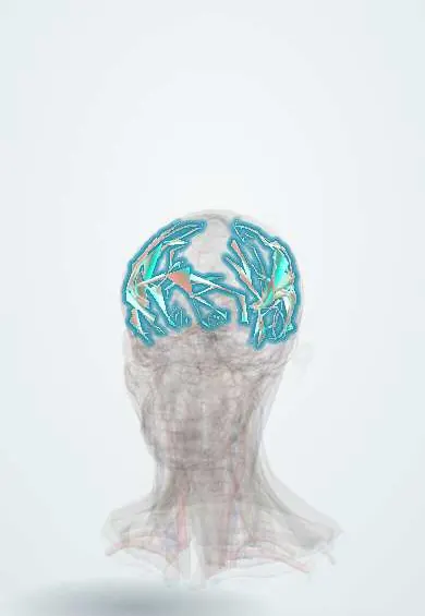
Brain arteriovenous malformation !
A tangle of abnormal blood vessels in the brain that links arteries directly to veins. It can bleed and is a common cause of brain hemorrhage in young people.
Updated 2026-11-01
Structures involved
Tap one to see what it is and open it in 3D.
- Middle cerebral artery
- Circle of Willis
- Anterior cerebral artery
- Posterior cerebral artery
- Internal carotid artery
- Superior sagittal sinus
- Transverse sinus
- Frontal lobe
- Parietal lobe
- Temporal lobe
- Occipital lobe
Why it happens
- Hemorrhage: petechiae, purpura, ecchymosis, hematoma, and what blood loss does
- Flow, pressure and resistance: Poiseuille and turbulent flow
- Blood vessels: artery, vein and capillary layers
What it is
A congenital vascular lesion in which arteries connect directly to veins through a tangle of abnormal vessels (the nidus) without a capillary bed. The high-flow, high-pressure shunt can rupture and cause intracerebral hemorrhage, or cause seizures and headache.
Who gets it
Found in roughly 1 in 1,000 to 2,000 people, often diagnosed between ages 20 and 40. It is one of the leading causes of non-traumatic intracerebral hemorrhage in children and young adults. Many are now found incidentally on brain imaging done for other reasons.
Causes
- Abnormal development of the vascular network during embryonic life; most are sporadic
- Hereditary hemorrhagic telangiectasia (Osler-Weber-Rendu), often with multiple malformations
- Somatic variants in vascular signalling genes (mainly KRAS) found in the vessel cells of many sporadic AVMs
Risk factors
- Previous rupture (the strongest predictor of new bleeding)
- Deep location or deep venous drainage only
- Associated aneurysms on feeding arteries
- Hereditary hemorrhagic telangiectasia
Pathophysiology
Without capillaries, arterial blood flows straight into thin-walled draining veins at high pressure. The veins dilate and may rupture, producing parenchymal hemorrhage, and less often subarachnoid or intraventricular bleeding. The high-flow shunt can also "steal" blood from surrounding brain, and the abnormal tissue irritates the cortex, causing seizures. Most occur in the cerebral hemispheres, often fed by branches of the middle cerebral artery.
Classification
- Spetzler-Martin grade (I to V): size of the nidus (<3, 3-6, >6 cm), eloquence of nearby brain, and presence of deep venous drainage; higher grades carry higher surgical risk
- Ruptured or unruptured
- Related lesions: cavernous malformation, developmental venous anomaly, dural arteriovenous fistula, vein of Galen malformation in infants
Symptoms and signs
- Sudden severe headache with vomiting, focal deficits or loss of consciousness when it bleeds
- Seizures (the second most common presentation)
- Chronic headache
- Progressive focal deficits from steal or mass effect
- Pulsating noise in the head (bruit) in some patients
- Heart failure and enlarged head in newborns with vein of Galen malformation
Diagnosis
- Clinical
- Suspected in a young person with intracerebral hemorrhage without hypertension, or with a first seizure. Neurological examination and Glasgow Coma Scale guide urgency.
- Tests
- Coagulation, blood count and type and screen before procedures. Electroencephalography in patients with seizures.
- Imaging
- Non-contrast CT shows acute hemorrhage. MRI and MR or CT angiography show the nidus and its relation to eloquent brain. Digital subtraction catheter angiography is the gold standard: it defines feeding arteries, the nidus, draining veins and associated aneurysms.
Differential diagnosis
- Hypertensive intracerebral hemorrhage
- Ruptured saccular aneurysm (subarachnoid hemorrhage)
- Cavernous malformation
- Hemorrhagic brain tumour (glioblastoma, metastasis)
- Cerebral venous thrombosis with hemorrhagic infarct
- Moyamoya disease
Goal of treatment
Stabilize acute hemorrhage, control seizures, and eliminate the malformation completely when the benefit outweighs the risk, to prevent future bleeding.
Treatment
- Acute
- Emergency care of intracerebral hemorrhage: airway, blood pressure control, reversal of anticoagulation, treatment of raised intracranial pressure and hydrocephalus, and evacuation of a large hematoma.
- Surgical
- Microsurgical resection of the nidus, preferred for low-grade (Spetzler-Martin I-II) accessible lesions; it offers immediate cure.
- Procedure
- Endovascular embolization with liquid agents, usually before surgery or radiosurgery to reduce flow, and stereotactic radiosurgery for small deep lesions, which closes the nidus over 1 to 3 years.
- Medical
- Antiseizure drugs for patients with seizures and analgesics for headache.
- Follow-up
- Conservative observation for many unruptured lesions, with blood pressure control, follow-up imaging after radiosurgery to confirm obliteration, and neurological checks after treatment.
Complications
- Intracerebral, intraventricular or subarachnoid hemorrhage
- Epilepsy
- Permanent neurological deficits
- Hydrocephalus
- Treatment complications: stroke, radiation injury, re-bleeding before complete obliteration
Prognosis
Unruptured malformations bleed at about 2 to 4% per year, higher after a first rupture. Each hemorrhage carries a significant risk of death or disability. Complete obliteration essentially eliminates the bleeding risk.
Test yourself
Think of the answer, then tap the question to see it.
What structure is missing between arteries and veins in an arteriovenous malformation?
The capillary bed; arteries drain directly into veins through a nidus.
What is the most common presentation of a brain AVM?
Intracerebral hemorrhage, followed by seizures.
Which study is the gold standard to define an AVM?
Digital subtraction catheter angiography.
Which three features make up the Spetzler-Martin grade?
Nidus size, eloquence of adjacent brain and presence of deep venous drainage.
Which hereditary disorder is associated with multiple brain AVMs?
Hereditary hemorrhagic telangiectasia (Osler-Weber-Rendu syndrome).
Related conditions
- Intracerebral haemorrhage
- Subarachnoid haemorrhage
- Epilepsy and seizures
- Status epilepticus
- Ischaemic stroke
- Migraine
- Raised intracranial pressure and brain herniation
- Cerebral venous sinus thrombosis
- Sturge–Weber syndrome
- Hydrocephalus
Educational information, not medical advice. For any symptom, see a health professional.
Sources: Cerebral arteriovenous malformation: MedlinePlus Medical Encyclopedia (MedlinePlus (U.S. National Library of Medicine), 2026).
Spotted a mistake?We are always improving for the community. Tell us what is wrong and our team will review it.Report a mistake
Representative model, for educational purposes only.