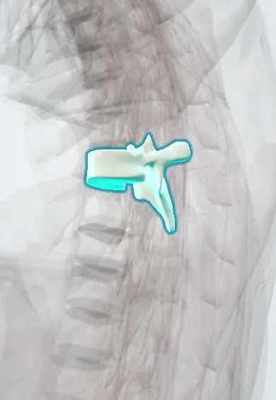
T6 vertebra !
T6 is the sixth thoracic (mid-back) vertebra; it has joints for the ribs and helps form the cage that protects the heart and lungs.
Official name: T6
Opens in the Durgical app, no sign-up needed.
What it is
A typical mid-back vertebra, with a heart-shaped body and small joints on each side for the ribs.
What it does
Holds up the trunk and, with the ribs and breastbone, forms the rib cage, which protects the heart and lungs. The ribs make this part of the spine stiff.
Key facts
- Joints with
- T5 above and T7 below (discs, facet joints); heads of ribs 6 and 7; tubercle of rib 6 (costotransverse joint)
- Landmarks
- Typical thoracic: heart-shaped body; costal demifacets above and below; transverse costal facets; long sloping spine; aortic impression on left side of body
Relations
- In front
- Esophagus moving slightly left, with the left atrium of the heart in front of it and the descending thoracic aorta against the left side of the body
- Lateral
- Heads of the sixth and seventh ribs; posterior intercostal arteries from the aorta wrap round the body; the azygos vein on the right
- Behind
- Trapezius and erector spinae; the inferior angle of the scapula lies about one level lower
- Contents
- Spinal cord in its narrowest and least well-supplied stretch
How to find it
Counted up one spine from T7, which lies level with the inferior angle of the scapula with the arms by the sides.
On imaging
In spinal cord infarction, axial T2 MRI may show two bright dots in the anterior horns ('owl's eyes'); diffusion imaging confirms an acute infarct. Lateral films show wedging if the body has collapsed.
Common problems
- Compression fractures from osteoporosis, sometimes after a minor fall or even a cough.
- A rounded upper back (kyphosis) from posture, wedge fractures or Scheuermann disease in teens.
- Scoliosis: a sideways curve with rotation, often starting in adolescence.
How it is diagnosed
X-rays for fractures and curves (scoliosis is measured with the Cobb angle). MRI tells new fractures from old ones. A bone density scan (DEXA) checks for osteoporosis.
Treatment
- Osteoporosis fractures: pain control, a short time in a brace, and treatment of the osteoporosis.
- Painful fractures that do not settle: kyphoplasty or vertebroplasty (cement injected into the bone).
- Large or progressive curves: bracing in growing children, sometimes fusion surgery.
Clinical notes
- Anterior spinal artery infarction (after aortic surgery, dissection or severe low blood pressure): sudden weak legs and loss of pain and temperature below the level, with position and vibration sense kept
- Autonomic dysreflexia shows as pounding headache, flushing above the injury and slow pulse; the trigger below the level, often bladder or bowel, is the key
- Neuraxial blocks are checked against dermatome landmarks: T6 at the xiphoid and T4 at the nipple; a caesarean section needs a block up to about T4, and a block that climbs higher also reaches the cardiac sympathetic fibres (T1–T4) and can slow the heart
Related conditions
Test yourself
Think of the answer, then tap the question to see it.
What is autonomic dysreflexia, a risk after spinal cord injury at T6 or above?
A dangerous surge in blood pressure triggered by a stimulus below the injury, often a full bladder: pounding headache, flushing above the injury and a slow pulse.
Why is the spinal cord at the level of the T6 vertebra prone to infarction?
It is a vascular watershed: feeder arteries are sparse between the upper thoracic supply and the artery of Adamkiewicz (about T4–T9).
What are the features of anterior spinal artery infarction of the mid-thoracic cord?
Sudden weak legs and loss of pain and temperature below the level, with position and vibration sense kept.
Which dermatome crosses the xiphoid process?
T6.
Educational information, not medical advice. For any symptom, see a health professional.
Its names
- Official Latin (TA2)
- vertebra thoracica VI TA2 1059
- Official Spanish (DeCS)
- Vértebra dorsal D6 DeCS D013904
- English (TA2)
- T6
Related
- T5–T6 disc
- T6–T7 disc
- Posterior intercostal arteries
- Anterior longitudinal ligament
- Rib-to-spine ligaments
- Endplates
- Mid-back facet joints
- Interspinous ligaments
Sources: Terminologia Anatomica (TA2); DeCS; Anatomy, Back, Thoracic Vertebrae.
Spotted a mistake?We are always improving for the community. Tell us what is wrong and our team will review it.Report a mistake
Representative model, for educational purposes only.