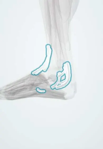
Ankle tendon sheaths !
The ankle tendon sheaths are slippery tubes around the tendons that cross the ankle, so they glide smoothly.
Official name: Anterior tarsal tendon sheaths
Opens in the Durgical app, no sign-up needed.
What it is
Slippery, fluid-filled tubes around the tendons that cross the ankle at the front, the inner side and the outer side.
What it does
Let the tendons glide smoothly under the retinacula and around the ankle bones.
Key facts
- Where
- Under retinacula: anterior (TA, EHL, EDL), medial (TP, FDL, FHL), lateral (common sheath of fibularis longus and brevis)
- Function
- Synovial sheaths that let tendons glide under retinacula and around malleoli; site of tenosynovitis
Relations
- In front
- Under the extensor retinacula, medial to lateral: tibialis anterior, extensor hallucis longus, extensor digitorum longus with fibularis tertius; the anterior tibial artery and deep fibular nerve lie between EHL and EDL
- Medial
- Under the flexor retinaculum (tarsal tunnel): tibialis posterior, flexor digitorum longus, the tibial nerve with the posterior tibial vessels, flexor hallucis longus
- Lateral
- Under the superior and inferior fibular retinacula: the common sheath of fibularis brevis (closer to the fibula) and fibularis longus; the sural nerve runs nearby
How to find it
Each tendon is found by resisting its action: tibialis anterior stands out in front with dorsiflexion and inversion; tibialis posterior is felt just behind the medial malleolus with resisted inversion; the fibular tendons are felt behind the lateral malleolus with resisted eversion. Sheath swelling follows the tendon's line.
Examination
- Single-heel-rise test: failure to rise on tiptoe on one leg, or the heel not swinging into varus, suggests tibialis posterior dysfunction
- Too-many-toes sign: seen from behind, more toes visible lateral to the heel on the affected side in adult acquired flatfoot
- Resisted eversion with the ankle dorsiflexed that makes the fibular tendons snap forward over the lateral malleolus shows subluxation from a torn superior fibular retinaculum
On imaging
Ultrasound, including dynamic scanning, shows sheath fluid, synovial thickening, tears and tendon subluxation; MRI shows tenosynovitis and longitudinal split tears. A little fluid in the flexor hallucis longus sheath can be normal because it may communicate with the ankle joint.
Common problems
- Tenosynovitis: painful swelling of a sheath from overuse, injury or inflammatory arthritis.
Clinical notes
- Fibularis brevis split tear: the tendon is squeezed between fibularis longus and the fibular groove
- Flexor hallucis longus tenosynovitis in ballet dancers, behind the medial malleolus, often with an os trigonum and posterior ankle impingement
- Tarsal tunnel syndrome: the tibial nerve is compressed under the flexor retinaculum, sometimes by tenosynovitis or a ganglion from a sheath
Anatomical variants
- Os peroneum, a sesamoid bone in the fibularis longus tendon near the cuboid, is common and can fracture or cause lateral foot pain
- An extra muscle, fibularis quartus, can crowd the fibular tendons behind the lateral malleolus
Memory aid
Behind the medial malleolus, front to back: "Tom, Dick And Very Nervous Harry" (Tibialis posterior, flexor Digitorum longus, Artery, Vein, Nerve, flexor Hallucis longus).
Related conditions
Test yourself
Think of the answer, then tap the question to see it.
What lies behind the medial malleolus, from front to back?
Tibialis posterior, flexor digitorum longus, posterior tibial artery and veins, tibial nerve, flexor hallucis longus.
How are the fibularis tendon sheaths arranged behind the lateral malleolus?
Fibularis longus and brevis share one sheath that splits in two lower down; the brevis lies against the bone.
Which tenosynovitis is an early stage of adult acquired flatfoot, and which test checks it?
Tibialis posterior tenosynovitis; the single-heel-rise test fails or the heel does not swing into varus.
Which tenosynovitis is typical of ballet dancers behind the medial malleolus?
Flexor hallucis longus tenosynovitis, often with an os trigonum and posterior ankle impingement.
Educational information, not medical advice. For any symptom, see a health professional.
Its names
- Official Latin (TA2)
- vaginae tarseae anteriores tendinum; vaginae tarseae tibiales tendinum; vaginae tarseae fibulares tendinum TA2 2755
- Official Spanish (DeCS)
- Vainas tendinosas tarsianas anteriores, tibiales y fibulares
- English (TA2)
- Anterior tarsal tendon sheaths
Related
Sources: Terminologia Anatomica (TA2); Anatomy, Bony Pelvis and Lower Limb: Foot and Ankle.
Spotted a mistake?We are always improving for the community. Tell us what is wrong and our team will review it.Report a mistake
Representative model, for educational purposes only.