Conditions · Surgery and trauma
Blunt traumatic aortic injury !
A tear of the aorta from a sudden stop, as in a crash or fall. It is often fatal; survivors need CT and usually a stent graft.
Updated 2026-11-01
Structures involved
Tap one to see what it is and open it in 3D.
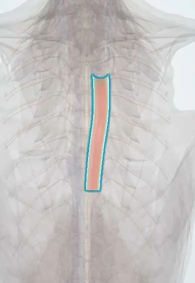
- Thoracic aorta
- Aortic arch
- Ligamentum arteriosum
- Ascending aorta
- Subclavian artery
- Esophagus
- Trachea
- Pulmonary trunk
Why it happens
- Hemorrhage: petechiae, purpura, ecchymosis, hematoma, and what blood loss does
- Blood vessels: artery, vein and capillary layers
What it is
Partial or full-thickness tear of the thoracic aorta caused by rapid deceleration, most often at the aortic isthmus just beyond the left subclavian artery, where the ligamentum arteriosum tethers the vessel. Most patients die at the scene; those who reach hospital have a contained injury that can still rupture.
Who gets it
It is a leading cause of death at the scene of high-speed crashes and falls. Survivors who reach hospital are a minority. In Colombia, motorcycle crashes, car crashes and falls from height are the usual mechanisms.
Causes
- High-speed motor vehicle or motorcycle crashes, especially frontal or side impacts
- Run-over pedestrians
- Falls from height
- Crush injuries of the chest
Risk factors
- High-energy deceleration mechanism
- No seatbelt or ejection from the vehicle
- Associated first or second rib, sternal or scapular fractures
- Older age (stiffer aorta)
Pathophysiology
During sudden deceleration the mobile aortic arch and heart keep moving while the descending aorta stays fixed to the spine, concentrating shear and bending forces at the isthmus. The tear runs from intima outward; if the adventitia and surrounding tissues hold, a contained haematoma or pseudoaneurysm forms, which can rupture at any time.
Classification
- Grade I: intimal tear
- Grade II: intramural haematoma
- Grade III: pseudoaneurysm
- Grade IV: rupture (free contrast extravasation)
Symptoms and signs
- Often no specific signs, masked by other injuries
- Chest or back (interscapular) pain
- Different blood pressure or pulses between the arms, or between arms and legs
- Hoarseness or difficulty swallowing from mediastinal haematoma
- Hypotension in rupture
Diagnosis
- Clinical
- Suspect it in every high-speed deceleration injury regardless of symptoms; the mechanism drives the work-up.
- Tests
- Blood group and crossmatch, haemoglobin, blood gas and lactate. Continuous arterial pressure monitoring in the resuscitation room.
- Imaging
- Chest X-ray as a screening adjunct (widened mediastinum and related signs). CT angiography of the chest is the diagnostic test of choice. Transoesophageal echocardiography in unstable patients who cannot go to CT.
Differential diagnosis
- Mediastinal haematoma from sternal or thoracic spine fracture
- Supine anteroposterior chest X-ray magnifying the mediastinum
- Blunt cardiac injury
- Spontaneous aortic dissection
- Great vessel branch injury
Goal of treatment
Prevent free rupture by lowering aortic wall stress, then repair the aorta at the safest time, usually with an endovascular stent graft.
Treatment
- Acute
- Anti-impulse therapy: lower heart rate first with a short-acting intravenous beta-blocker, then blood pressure with a vasodilator if needed, as other injuries allow; analgesia.
- Procedure
- Thoracic endovascular aortic repair (TEVAR) is preferred over open surgery when anatomy permits.
- Surgical
- Open surgical repair with graft interposition when endovascular repair is not possible.
- Conservative
- Grade I intimal tears are often managed with blood pressure control and follow-up imaging; repair can be delayed for higher grades while life-threatening injuries are treated, if pressure is controlled.
- Follow-up
- Nursing priorities: continuous arterial pressure and heart rate monitoring with set targets, bilateral arm pressures, limb pulses, neurological checks for paraplegia after repair, and pain control.
Complications
- Free rupture and death
- Paraplegia from spinal cord ischaemia after repair
- Stent graft complications: endoleak, migration, left subclavian coverage
- Stroke
- Pseudoaneurysm formation if untreated
Prognosis
Most victims die before reaching hospital. Among those who arrive alive, rupture risk is highest in the first hours, and endovascular repair with good pressure control has lowered mortality and paraplegia compared with open surgery.
Prevention
- Seatbelts and airbags
- Speed control
- Motorcycle safety
Test yourself
Think of the answer, then tap the question to see it.
Where does blunt traumatic aortic injury most often occur?
At the aortic isthmus, just distal to the left subclavian artery, where the ligamentum arteriosum tethers the aorta.
What is the classic chest X-ray sign of blunt aortic injury?
A widened mediastinum, often with loss of the aortic knob, tracheal or nasogastric tube deviation to the right, or a left apical cap.
What test confirms the diagnosis in a stable patient?
CT angiography of the chest.
What is the preferred repair for most patients?
Thoracic endovascular aortic repair (stent graft), after heart rate and blood pressure control.
Related conditions
- Major trauma (polytrauma): ATLS primary survey
- Aortic dissection
- Rib fractures
- Flail chest
- Haemothorax
- Blunt cardiac injury
- Cardiac tamponade
- Hypovolaemic shock
- Pneumothorax
Educational information, not medical advice. For any symptom, see a health professional.
Sources: Evaluation and management of blunt traumatic aortic injury: a practice management guideline from the Eastern Association for the Surgery of Trauma (Journal of Trauma Nursing (PubMed), 2015).
Spotted a mistake?We are always improving for the community. Tell us what is wrong and our team will review it.Report a mistake
Representative model, for educational purposes only.